
Page 1166 - Hall et al (2015) Principles of Critical Care-McGraw-Hill
P. 1166
CHAPTER 86: Intracranial Pressure: Monitoring and Management 805
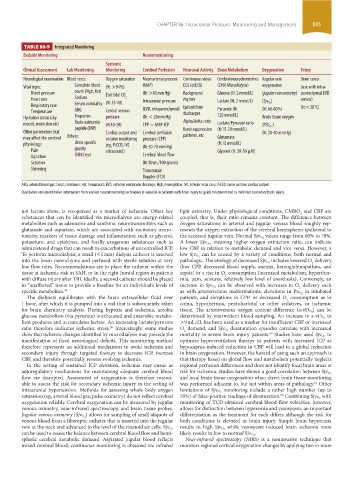
TABLE 86-9 Integrated Monitoring
Bedside Monitoring Neuromonitoring
Systemic
Clinical Assessment lab Monitoring Monitoring Cerebral Perfusion Neuronal Activity Brain Metabolism Oxygenation Temp
Neurological examination Blood tests: Oxygen saturation Mean arterial pressure Continuous video Cerebral neurochemistries Jugular vein Brain temp
Vital signs: Complete blood (N: >94%) (MAP) EEG (cvEEG) (CMA Microdialysis) oxygenation (use with intra-
Blood pressure count (Hgb, Hct) End tidal CO (N: >80 mm Hg) Background Glucose (N: 2 mmol/L) (jugular vein oximetry) parenchymal EVD
Heart rate Sodium 2 Intracranial pressure rhythm Lactate (N: 2 mmol/L) ) sensor)
Respiratory rate Serum osmolality (N: 35-40) Epileptiform (Sjv O 2 (N:<38 °C)
Temperature ABG Central venous (EVD, intraparenchymal) discharges Pyruvate (N: (N: 60-80%)
Hydration status (dry Troponins pressure (N: < 20 mm Hg) 120 mmol/L) Brain tissue oxygen
mouth, moist skin etc) Brain-natriuretic (N:10-20) CPP = MAP-ICP Alpha/delta ratio Lactate/Pyruvate ratio )
peptide (BNP) Burst suppression (N:15-20 mmol/L) (PBt O 2
Other parameters that Cardiac output and Cerebral perfusion (N: 20-40 mm Hg)
may affect the cerebral Others: volume monitoring pressure (CPP) patterns, etc Glutamate
physiology: Urine specific (eg, PiCCO, IVC (N: 50-70 mm Hg) (N:10 mmol/L)
Pain gravity ultrasound) Glycerol (N: 20-50 µM)
Agitation BUN:Creat Cerebral blood flow
Sedation (N: 50 mL/100 g/min)
Shivering Transcranial
Doppler (TCD)
ABG, arterial blood gas; Creat, creatinine; Hct, hematocrit; EVD, external ventricular drainage; Hgb, hemoglobin; IVC, inferior vena cava; PiCCO, pulse contour cardiac output.
Qualitative and quantitative information from various neuromonitoring techniques is valuable in patients with brain injury to guide treatment and to minimize secondary brain injury.
not lactate alone, is recognized as a marker of ischemia. Other key light oximetry. Under physiological conditions, CMRO and CBF are
2
substances that can be identified via microdialysis are energy-related coupled, that is, their ratio remains constant. The difference between
metabolites such as adenosine and xanthine, neurotransmitters such as oxygen saturations in arterial and jugular venous blood roughly rep-
glutamate and aspartate, which are associated with excitatory neuro- resents the oxygen extraction of the cerebral hemispheres ipsilateral to
toxicity, markers of tissue damage and inflammation such as glycerol, the accessed jugular vein. Normal Sjv O 2 values range from 60% to 70%.
potassium, and cytokines, and finally exogenous substances such as A lower Sjv O 2 , meaning higher oxygen extraction ratio, can indicate
administered drugs that can result in exacerbations of uncontrolled ICP. low CBF in relation to metabolic demand and vice versa. However, a
To perform microdialysis, a small (<1 mm) dialysis catheter is inserted low Sjv O 2 can be caused by a variety of conditions, both normal and
into the brain parenchyma and perfused with sterile solution at very pathologic. The etiology of decreased Sjv O 2 includes lowered O delivery
2
low flow rates. Recommendations are to place the catheter within the (low CPP, decreased blood supply, anemia, hemoglobinopathies, and
tissue at ischemic risk in SAH, or in the right frontal region in patients sepsis) or a rise in O consumption (increased metabolism, hyperther-
2
with diffuse injury after TBI. Ideally, a second catheter should be placed mia, pain, seizures, relatively low level of anesthesia). Conversely, an
in “unaffected” tissue to provide a baseline for an individual’s levels of increase in Sjv O 2 can be observed with increases in O delivery such
2
specific metabolites. 58 as with arteriovenous malformations, elevations in Pa O 2 in intubated
The dialysate equilibrates with the brain extracellular fluid over patients, and elevations in CPP, or decreased O consumption as in
2
1 hour, after which it is pumped into a vial that is subsequently taken coma, hypothermia, pentobarbital or other sedatives, or ischemic
for brain chemistry analysis. During hypoxia and ischemia, aerobic tissue. The arteriovenous oxygen content difference (a-vD O 2 ) can be
glucose metabolism (via pyruvate) is exhausted and anaerobic metabo- determined by intermittent blood sampling. An increase in a-vD O 2 to
lism produces and accumulates lactate. An increasing lactate/pyruvate >9 mL/dL has been used as a marker for insufficient CBF or increased
ratio therefore indicates ischemic stress. Interestingly, some studies O demand, and Sjv O 2 desaturation episodes correlate with increased
59
2
show that ischemic changes identified by microdialysis may precede the mortality in severe brain injury patients. Studies have used Sjv O 2 to
60
manifestation of fixed neurological deficits. This monitoring method optimize hyperventilation therapy in patients with increased ICP as
therefore represents an additional mechanism to avoid ischemia and hypocapnia-induced reduction in CBF will lead to a global reduction
secondary injury through targeted therapy to decrease ICP, increase in brain oxygenation. However, the hazard of using such an approach is
CBF, and therefore potentially reverse evolving ischemia. that therapy based on global flow and metabolism potentially neglects
In the setting of sustained ICP elevation, ischemia may ensue as regional perfusion differences and does not identify focal brain areas at
autoregulatory mechanisms for maintaining adequate cerebral blood risk for ischemia. Studies have shown a good correlation between Sjv O 2
flow are disrupted. Assessment of oxygenation is therefore reason- and local brain tissue oxygenation when direct brain tissue monitoring
able to assess the risk for secondary ischemic injury in the setting of was performed adjacent to, but not within areas of pathology. Other
61
intracranial hypertension. Methods for assessing whole body oxygen limitations of Sjv O 2 monitoring include a rather high number (up to
saturation (eg, arterial blood gas, pulse oximetry) do not reflect cerebral 50%) of false-positive readings of desaturation. Combining Sjv O 2 with
62
oxygenation reliably. Cerebral oxygenation can be measured by jugular monitoring of TCD-obtained cerebral blood flow velocities, however,
venous oximetry, near-infrared spectroscopy, and brain tissue probes. allows for distinction between hyperemia and vasospasm, an important
) allows for sampling of small aliquots of differentiation as the treatment for each differs although the risk for
Jugular venous oximetry (Sjv O 2
venous blood from a fiberoptic catheter that is inserted into the jugular both conditions is elevated in brain injury. Simple brain hyperemia
while vasospasm-induced brain ischemia more
vein at the neck and advanced to the level of the mastoid air cells. Sjv O 2 results in high Sjv O 2
can be used to assess the balance between cerebral blood flow and hemi- likely results in low to normal Sjv O 2 .
spheric cerebral metabolic demand. Aspirated jugular blood reflects Near-infrared spectroscopy (NIRS) is a noninvasive technique that
mixed cerebral blood; continuous monitoring is obtained via infrared monitors regional cortical oxygenation changes by applying two or more
section06.indd 805 1/23/2015 12:56:05 PM