
Page 1164 - Hall et al (2015) Principles of Critical Care-McGraw-Hill
P. 1164
CHAPTER 86: Intracranial Pressure: Monitoring and Management 803
CSF. Placement of these devices, however, is associated with morbidity abnormal ICP waveforms can be helpful in predicting and preventing
as with any invasive device. There is a risk of intracranial or intraven- malignant elevations in ICP as the noncompliant brain is often intolerant
tricular hemorrhage at the insertion site as well as a risk of infection that of additional volume within the intracranial contents without brisk eleva-
can range from 6% to 11%. 39-41 Infection risk increases over time and the tions in ICP (Fig. 86-4, Area B). For example, when a patient has fulminant
consequences can be associated with significant morbidity and mortality hepatic failure, observation of these subtle changes in the ICP waveform can
if ventriculitis develops. signal the evolution of brain swelling before it is clinically apparent or before
■ MONITORING OF ABNORMAL ICP the ICP has increased.
ICP elevation can also be predicted imperfectly through brain imag-
The clinical course of ICP elevation is variable and largely determined by ing studies. Nevertheless, when the ICP from any monitor type is either
the etiology of the intracranial hypertension. Certain causes of elevated much lower or higher than expected based on clinical or radiographic
ICP are more progressive than others. In this setting of TBI, intracra- findings, steps should be taken to determine the accuracy of the monitor.
nial hypertension often occurs early, within 72 hours. It may, however, At times this may require rezeroing or replacing the monitor, and rarely
develop late or follow a bimodal pattern, and as many as 25% of patients inserting a new monitor in a new position or location. When there is
experience their highest ICP after 5 days. Therefore, it is important to significant discrepancy between the ICP and clinical presentation and
recall that ICP elevations show a temporal heterogeneity within the first imaging results, replacement or rezeroing of the monitor should be
2 weeks of injury, creating another strong argument for continuous ICP considered. It is important to realize that ICP is not homogeneous due
monitoring. to compartmentalization of intracranial structures.
form temporally associated with the systemic blood pressure. Certain ■ MONITORING CEREBRAL AUTOREGULATION
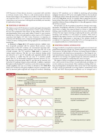
As illustrated in Figure 86-3, ICP monitors provide a ballistic wave-
characteristics of the ICP waveform can communicate important infor- Testing of cerebral autoregulation requires the application of a timed and
mation about an evolving cerebral process, even when the ICP is not graded hemodynamic stimulus (eg, carotid artery compression, negative
critically elevated. The ICP waveform (Table 86-8) is comprised of body pressure, tilt table, thigh cuff release, or pharmacological change in
various smaller subwaves. The first two waves observed during systole blood pressure) with simultaneous measurement of the change in cere-
are referred to as P1 and P2. Normally, each sequential subwave of the bral hemodynamic response. Since clinical and practical reasons make it
ICP waveform is smaller than the one prior. P1 is therefore usually difficult for such testing to be applied frequently, alternative methods of
greater than P2. As intracranial compliance decreases, however, P2 usu- continuous autoregulation monitoring have been developed.
ally increases to become greater than P1, and this can be observed even The degree of intact autoregulatory mechanisms can fluctuate widely
before the ICP reading increases to abnormal levels. In addition, respiratory over a short period of time in brain injured individuals, providing a rea-
fluctuations in the baseline ICP waveform usually reflect a decrease in intra- son for continuous measurements. Most continuous testing of the func-
cranial compliance and can occur before an elevation in ICP. Table 86-8 tion of autoregulatory mechanisms relies on the beat-to-beat responses
depicts ICP waveforms with good compliance and poor compliance, indi- of CBF to other spontaneously occurring, rhythmic measurements such
cated by increased pulse amplitude and dampened waveform. Identifying as CPP and MAP. Hemodynamic oscillations can be averaged over a
TABLE 86-8 ICP Waveform Analysis
Compliance ICP Waveform Conditions Related cerebral physiology
P1: percussion wave–transmitting through the cerebral arterial tree to the choroid (examples)
plexus (ventricles)
P2: tidal wave–compliance; early impairment in cerebral vasomotor paralysis,
brain swelling, etc; reflects the venous compartment and its
normal amplitude is 80% of P1
P3: dichrotic wave–reflects the aortic valve closure
Normal P1 Normal ICP, normal brain Three peaks of decreasing height.
compliance P2 compliance P1 generally with a sharp peak and a fairly
P3 constant amplitude. P2 is more variable and
ends at the dichrotic notch. P3 follows the
dichrotic notch and is not discernable at times
Reduced P1 P2 Severe arterial Decrease mean ICP; decrease ICP waveform
compliance P3 hypotension; amplitude P2 with little change in P1
hyperventilation
Increased P1 P2 Rapidly expanding Increases mean ICP
amplitude P3 mass lesion; severe Increases ICP amplitude, mainly P2 and P3
arterial hypertension; Rounding of ICP waveform due to increase in
severe hypercapnia and/or later waveform components
hypoxia; jugular vein
compression
Dampened Individuals with ICP waveform dampened and low in amplitude
waveform craniectomy or open
skull (TBI)
ICP waveform analysis of the common pressure abnormalities offers important bedside information about intracranial pressure dynamics helpful in clinical decision-making. ICP, intracranial pressure; TBI, traumatic brain injury
section06.indd 803 1/23/2015 12:56:05 PM