
Page 1161 - Hall et al (2015) Principles of Critical Care-McGraw-Hill
P. 1161
800 PART 6: Neurologic Disorders
disc. Paton lines, defined as circumferential peripapillary retinal folds
caused by inward buckling of the swollen disc, are another manifestation
of optic nerve edema secondary to raised ICP.
In general, pupillary dilation is seen as a result of third nerve compres-
sion identified by pupillary dilation with ptosis, sparing abduction, and
intorsion-depression. A third cranial nerve palsy can be seen as a result
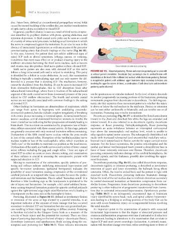
of a variety of intracranial processes, including uncal herniation in which Decorticate or flexor posturing
the nerve is compressed against the tentorium. It can also be seen in the
absence of intracranial hypertension as with aneurysms of the posterior
communicating artery that directly impinge on the nerve (Fig. 86-1B).
In this case, however, the patient does not exhibit additional signs of
raised ICP such as altered sensorium, lethargy, nausea, or vomiting.
Conditions that exert mass effect on or produce shearing injury to the
midbrain structures harboring the third nerve nucleus, such as tumors
and trauma, may also produce third nerve palsies without elevated ICP. Decerebrate or extensor posturing
Sixth nerve palsies are also seen with elevated ICP as this nerve
follows a long intracranial path and is subject to stretching. This palsy FIGURE 86-15. Abnormal posturing. Flexion and extension posturing can be seen with
is identified by a deficit in ocular abduction. As such, this examination or without patient stimulation. Decorticate (top) posturing is due to cerebral lesions with
finding is typically a nonlocalizing sign and can only narrow the dif- disinhibition at the level of the midbrain (red nucleus) while decerebrate posturing (bottom)
ferential to a process that is elevating ICP. The mechanism, however, is recognized in patients with additional upper brainstem injury secondary to lesions also
cannot be elucidated from this finding. (It can be seen in increased ICP involving the upper brainstem. At times, a combination of both decorticate and decerebrate
from obstructive hydrocephalus, that is, CSF absorption block after posturing can be observed.
subarachnoid hemorrhage, where there is traction of the subarachnoid
segment of the sixth cranial nerve from downward pressure of the pons can be spontaneous or stimulus induced. As the level of injury descends
related to CSF outflow obstruction.) The fourth and seventh cranial to involve progressively increasing portions of the brainstem, posturing
nerves are not typically associated with common findings in the setting movements progress from decorticate to decerebrate. The defining ana-
of elevated ICP. tomic site that separates these movement patterns is whether the injury
Other findings in herniation are abnormalities of respiration, which is above or below the red nucleus in the midbrain. Flexion or extension
can range from apnea to hyperpnea with undulating crescendo- can be seen either unilaterally or bilaterally, and can involve one or all
decrescendo patterns, but also include complete irregularity of breathing extremities. Posturing may even be intermittent.
with erratic pauses increasing to terminal apnea. As intracranial hyper- Decorticate posturing (Fig. 86-15) is identified by flexed arms drawn
tension escalates, central downward herniation worsens to involve the inward to the chest and clenched fists while the legs are extended and
lower cranial nerves indicating further progression ultimately leading to rotated inward. It is also referred to as decorticate rigidity, decorticate
brain death. This is evident on examination by progressive loss of brain- response, or flexor posturing. Pathophysiologically, flexor posturing
stem reflexes associated with these cranial nerves. At this stage, patients represents an acute imbalance between an interrupted corticospinal
are generally comatose with only minimal brainstem reflexes remaining. tract above the mesencephalic red nucleus level, which is unable to
Dysfunction of the fifth cranial nerve nucleus within the pons results relay signals to spinal motor neurons. The subsequently disinhibited red
in loss of the corneal reflex. Disruption of the vestibulocochlear nerve nuclei with increased rubrospinal (and uncontrolled medullary reticu-
nucleus results in loss of the oculocephalic reflex, which manifests as lospinal tract) output leads to flexion activity for upper cervical motor
“doll’s eyes” or the inability to maintain eye position as the head moves. neurons. For the lower extremities, the pontine reticulospinal and the
Dysfunction of the ninth and tenth nerves results in loss of lower cranial medial and lateral vestibulospinal tracts present a disinhibition bias in
nerve reflexes including the gag and cough reflex. These are signs of favor of lower extremity extension over flexion. Therefore, decorticate
raised ICP in either an acute or more chronic setting, and examination posturing commonly indicates damage of the cerebral hemispheres, the
of these reflexes is crucial in assessing the unresponsive patient with internal capsule, and the thalamus, possibly also involving the upper-
suspected elevation in ICP. most brainstem.
Moving to examination of the extremities, specific patterns of pos- Decerebrate posturing (Fig. 86-15), also called decerebrate response,
turing and reflexes indicate underlying intracranial hypertension and decerebrate rigidity, or extensor posturing, is described as involuntary
specific herniation syndromes. Ipsilateral hemiparesis indicates the extension of upper and lower extremities, the hallmark being elbow
possibility of uncal herniation causing compression of the contralateral extension. Often, the head is arched back and the patient is rigid with
cerebral peduncle as temporal lobe tissue extrudes between the ipsilat- clenched teeth. Decerebrate posturing indicates brainstem damage
eral tentorium and the brainstem. This is called “Kernohan notch”. It is below the level of the red nucleus due to midbrain distortion secondary
therefore a false localizing sign as the hemiparesis is ipsilateral to the to central downward herniation or brainstem compression from cer-
site of injury and herniation. For example, a left-sided subdural hema- ebellar lesions. Progression from decorticate posturing to decerebrate
toma causing temporal herniation pushes the opposite cerebral peduncle posturing is often indicative of progressive transtentorial brain hernia-
against the right tentorial edge (right-sided Kernohan notch), leading to tion due to untreated intracranial hypertension. Opisthotonic postur-
a hemiparesis on the same side as the hemispheric lesion. ing (Table 86-5) is an infrequently encountered sign of brainstem
Posturing, illustrated in Figure 86-15, defined as involuntary flexion injury identified by severe head, neck, and spinal column hyperexten-
or extension of the arms or legs elicited by a painful stimulus, is an sion leading to a bridging or arching position of the body that can be
important indicator of the amount of brain damage that has occurred seen with severe brainstem injury or extrapyramidal lesions involving
secondary to elevated ICP and herniation but also any generalized pro- the axial muscles.
cess resulting in brain injury (eg, shearing or diffuse anoxia). Posturing An important point in the examination of patients with suspected
is therefore included in the Glasgow Coma Scale as a measure of the intracranial hypertension is that the degree of abnormal pressure and
severity of brain injury and the potential for recovery. There are three craniocaudal herniation progresses over time if untreated or if refractory
types of posturing depending on the level of injury—decorticate (flexor), to treatment, leading to alterations in the examination that correlate to
decerebrate (extensor), and opisthotonos (body arching along the cra- higher ICP and more severe neurologic dysfunction. A patient’s exami-
niospinal axis) posturing (see Table 86-5). These reflexive movements nation will therefore change over time, either involving more or fewer of
section06.indd 800 1/23/2015 12:56:02 PM