
Page 132 - Clinical Anatomy
P. 132
ECA2 7/18/06 6:43 PM Page 117
The male genital organs 117
between these three ducts. In benign prostatic hypertrophy, (but not in the
normal prostate), a shallow posterior median groove (which can be felt on
rectal examination) further divides the prostate into left and right lobes.
Anterior to the urethra, the prostate consists of a narrow isthmus only.
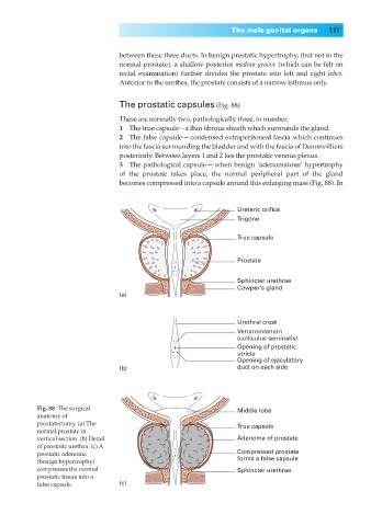
The prostatic capsules (Fig. 88)
These are normally two, pathologically three, in number.
1◊◊The true capsule—a thin fibrous sheath which surrounds the gland.
2◊◊The false capsule — condensed extraperitoneal fascia which continues
into the fascia surrounding the bladder and with the fascia of Denonvilliers
posteriorly. Between layers 1 and 2 lies the prostatic venous plexus.
3◊◊The pathological capsule — when benign ‘adenomatous’ hypertrophy
of the prostate takes place, the normal peripheral part of the gland
becomes compressed into a capsule around this enlarging mass (Fig. 88). In
Ureteric orifice
Trigone
True capsule
Prostate
Sphincter urethrae
Cowper's gland
(a)
Urethral crest
Verumontanum
(colliculus seminalis)
Opening of prostatic
utricle
Opening of ejaculatory
(b) duct on each side
Fig. 88◊The surgical Middle lobe
anatomy of
prostatectomy. (a) The True capsule
normal prostate in
vertical section. (b) Detail Adenoma of prostate
of prostatic urethra. (c) A
prostatic adenoma Compressed prostate
(benign hypertrophy) forms a false capsule
compresses the normal Sphincter urethrae
prostatic tissue into a
false capsule. (c)