
Page 127 - Clinical Anatomy
P. 127
ECA2 7/18/06 6:43 PM Page 112
112 The abdomen and pelvis
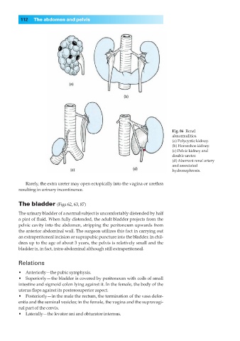
Fig. 86◊Renal
abnormalities.
(a) Polycystic kidney.
(b) Horseshoe kidney.
(c) Pelvic kidney and
double ureter.
(d) Aberrant renal artery
and associated
hydronephrosis.
Rarely, the extra ureter may open ectopically into the vagina or urethra
resulting in urinary incontinence.
The bladder (Figs 62, 63, 87)
The urinary bladder of a normal subject is uncomfortably distended by half
a pint of fluid. When fully distended, the adult bladder projects from the
pelvic cavity into the abdomen, stripping the peritoneum upwards from
the anterior abdominal wall. The surgeon utilizes this fact in carrying out
an extraperitoneal incision or suprapubic puncture into the bladder. In chil-
dren up to the age of about 3 years, the pelvis is relatively small and the
bladder is, in fact, intra-abdominal although still extraperitoneal.
Relations
•◊◊Anteriorly—the pubic symphysis.
•◊◊Superiorly — the bladder is covered by peritoneum with coils of small
intestine and sigmoid colon lying against it. In the female, the body of the
uterus flops against its posterosuperior aspect.
•◊◊Posteriorly— in the male the rectum, the termination of the vasa defer-
entia and the seminal vesicles; in the female, the vagina and the supravagi-
nal part of the cervix.
•◊◊Laterally—the levator ani and obturator internus.