
Page 138 - Clinical Anatomy
P. 138
ECA2 7/18/06 6:43 PM Page 123
The male genital organs 123
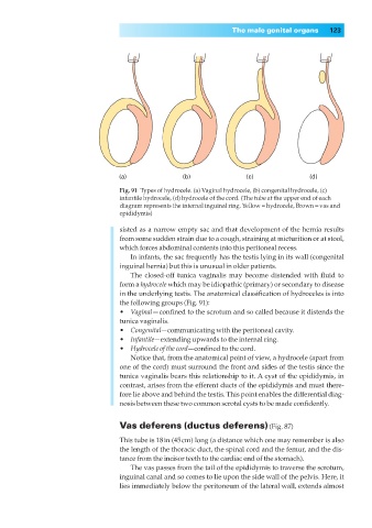
(a) (b) (c) (d)
Fig. 91◊Types of hydrocele. (a) Vaginal hydrocele, (b) congenital hydrocele, (c)
infantile hydrocele, (d) hydrocele of the cord. (The tube at the upper end of each
diagram represents the internal inguinal ring. Yellow = hydrocele, Brown = vas and
epididymis)
sisted as a narrow empty sac and that development of the hernia results
from some sudden strain due to a cough, straining at micturition or at stool,
which forces abdominal contents into this peritoneal recess.
In infants, the sac frequently has the testis lying in its wall (congenital
inguinal hernia) but this is unusual in older patients.
The closed-off tunica vaginalis may become distended with fluid to
form a hydrocele which may be idiopathic (primary) or secondary to disease
in the underlying testis. The anatomical classification of hydroceles is into
the following groups (Fig. 91):
•◊◊Vaginal— confined to the scrotum and so called because it distends the
tunica vaginalis.
•◊◊Congenital—communicating with the peritoneal cavity.
•◊◊Infantile—extending upwards to the internal ring.
•◊◊Hydrocele of the cord—confined to the cord.
Notice that, from the anatomical point of view, a hydrocele (apart from
one of the cord) must surround the front and sides of the testis since the
tunica vaginalis bears this relationship to it. A cyst of the epididymis, in
contrast, arises from the efferent ducts of the epididymis and must there-
fore lie above and behind the testis. This point enables the differential diag-
nosis between these two common scrotal cysts to be made confidently.
Vas deferens (ductus deferens) (Fig. 87)
This tube is 18in (45cm) long (a distance which one may remember is also
the length of the thoracic duct, the spinal cord and the femur, and the dis-
tance from the incisor teeth to the cardiac end of the stomach).
The vas passes from the tail of the epididymis to traverse the scrotum,
inguinal canal and so comes to lie upon the side wall of the pelvis. Here, it
lies immediately below the peritoneum of the lateral wall, extends almost