
Page 143 - Clinical Anatomy
P. 143
ECA2 7/18/06 6:43 PM Page 128
128 The abdomen and pelvis
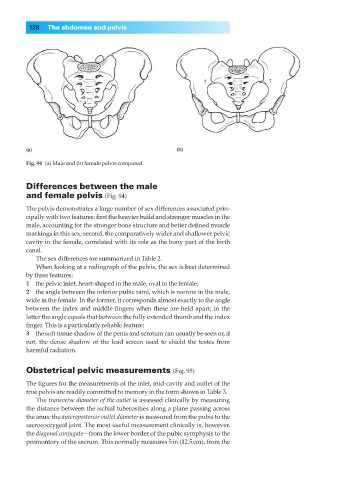
Fig. 94 (a) Male and (b) female pelvis compared.
Differences between the male
and female pelvis (Fig. 94)
The pelvis demonstrates a large number of sex differences associated prin-
cipally with two features: first the heavier build and stronger muscles in the
male, accounting for the stronger bone structure and better defined muscle
markings in this sex; second, the comparatively wider and shallower pelvic
cavity in the female, correlated with its role as the bony part of the birth
canal.
The sex differences are summarized in Table 2.
When looking at a radiograph of the pelvis, the sex is best determined
by three features:
1◊◊the pelvic inlet, heart-shaped in the male, oval in the female;
2◊◊the angle between the inferior pubic rami, which is narrow in the male,
wide in the female. In the former, it corresponds almost exactly to the angle
between the index and middle fingers when these are held apart; in the
latter the angle equals that between the fully extended thumb and the index
finger. This is a particularly reliable feature;
3◊◊the soft tissue shadow of the penis and scrotum can usually be seen or, if
not, the dense shadow of the lead screen used to shield the testes from
harmful radiation.
Obstetrical pelvic measurements (Fig. 95)
The figures for the measurements of the inlet, mid-cavity and outlet of the
true pelvis are readily committed to memory in the form shown in Table 3.
The transverse diameter of the outlet is assessed clinically by measuring
the distance between the ischial tuberosities along a plane passing across
the anus; the anteroposterior outlet diameter is measured from the pubis to the
sacrococcygeal joint. The most useful measurement clinically is, however,
the diagonal conjugate—from the lower border of the pubic symphysis to the
promontory of the sacrum. This normally measures 5in (12.5cm); from the