
Page 140 - Clinical Anatomy
P. 140
ECA2 7/18/06 6:43 PM Page 125
The bony and ligamentous pelvis 125
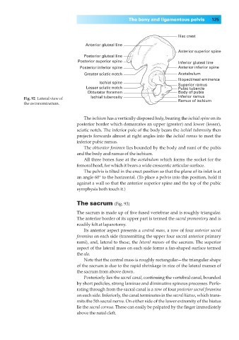
Iliac crest
Anterior gluteal line
Anterior superior spine
Posterior gluteal line
Posterior superior spine Inferior gluteal line
Posterior inferior spine Anterior inferior spine
Greater sciatic notch Acetabulum
Iliopectineal eminence
Ischial spine
Superior ramus
Lesser sciatic notch Pubic tubercle
Obturator foramen Body of pubis
Fig. 92 Lateral view of Ischial tuberosity Inferior ramus
Ramus of ischium
the os innominatum.
The ischium has a vertically disposed body, bearing the ischial spine on its
posterior border which demarcates an upper (greater) and lower (lesser),
sciatic notch. The inferior pole of the body bears the ischial tuberosity then
projects forwards almost at right angles into the ischial ramus to meet the
inferior pubic ramus.
The obturator foramen lies bounded by the body and rami of the pubis
and the body and ramus of the ischium.
All three bones fuse at the acetabulum which forms the socket for the
femoral head, for which it bears a wide crescentic articular surface.
The pelvis is tilted in the erect position so that the plane of its inlet is at
an angle 60° to the horizontal. (To place a pelvis into this position, hold it
against a wall so that the anterior superior spine and the top of the pubic
symphysis both touch it.)
The sacrum (Fig. 93)
The sacrum is made up of five fused vertebrae and is roughly triangular.
The anterior border of its upper part is termed the sacral promontory and is
readily felt at laparotomy.
Its anterior aspect presents a central mass, a row of four anterior sacral
foramina on each side (transmitting the upper four sacral anterior primary
rami), and, lateral to these, the lateral masses of the sacrum. The superior
aspect of the lateral mass on each side forms a fan-shaped surface termed
the ala.
Note that the central mass is roughly rectangular—the triangular shape
of the sacrum is due to the rapid shrinkage in size of the lateral masses of
the sacrum from above down.
Posteriorly lies the sacral canal, continuing the vertebral canal, bounded
by short pedicles, strong laminae and diminutive spinous processes. Perfo-
rating through from the sacral canal is a row of four posterior sacral foramina
on each side. Inferiorly, the canal terminates in the sacral hiatus, which trans-
mits the 5th sacral nerve. On either side of the lower extremity of the hiatus
lie the sacral cornua. These can easily be palpated by the finger immediately
above the natal cleft.