
Page 217 - Clinical Anatomy
P. 217
ECA3 7/18/06 6:45 PM Page 202
202 The upper limb
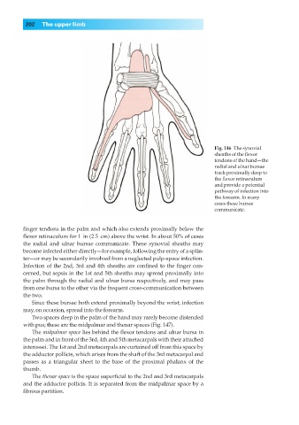
Fig. 146◊The synovial
sheaths of the flexor
tendons of the hand—the
radial and ulnar bursae
track proximally deep to
the flexor retinaculum
and provide a potential
pathway of infection into
the forearm. In many
cases these bursae
communicate.
finger tendons in the palm and which also extends proximally below the
flexor retinaculum for 1 | in (2.5 | cm) above the wrist. In about 50% of cases
the radial and ulnar bursae communicate. These synovial sheaths may
become infected either directly—for example, following the entry of a splin-
ter—or may be secondarily involved from a neglected pulp-space infection.
Infection of the 2nd, 3rd and 4th sheaths are confined to the finger con-
cerned, but sepsis in the 1st and 5th sheaths may spread proximally into
the palm through the radial and ulnar bursa respectively, and may pass
from one bursa to the other via the frequent cross-communication between
the two.
Since these bursae both extend proximally beyond the wrist, infection
may, on occasion, spread into the forearm.
Two spaces deep in the palm of the hand may rarely become distended
with pus; these are the midpalmar and thenar spaces (Fig. 147).
The midpalmar space lies behind the flexor tendons and ulnar bursa in
the palm and in front of the 3rd, 4th and 5th metacarpals with their attached
interossei. The 1st and 2nd metacarpals are curtained off from this space by
the adductor pollicis, which arises from the shaft of the 3rd metacarpal and
passes as a triangular sheet to the base of the proximal phalanx of the
thumb.
The thenar space is the space superficial to the 2nd and 3rd metacarpals
and the adductor pollicis. It is separated from the midpalmar space by a
fibrous partition.