
Page 312 - Clinical Anatomy
P. 312
ECA5 7/18/06 6:50 PM Page 297
The major arteries of the head and neck 297
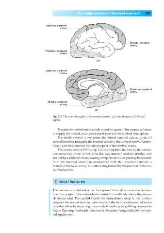
Fig. 211◊The arterial supply of the cerebral cortex. (a) Lateral aspect. (b) Medial
aspect.
The anterior cerebral artery winds round the genu of the corpus callosum
to supply the medial and superolateral aspect of the cerebral hemisphere.
The middle cerebral artery enters the lateral cerebral sulcus, gives off
central branches to supply the internal capsule (‘the artery of cerebral haemor-
rhage’) and feeds most of the lateral aspect of the cerebral cortex.
The arterial circle of Willis (Fig. 212) is completed in front by the anterior
communicating artery, which links the two anterior cerebral arteries, and
behind by a posterior communicating artery on each side, passing backwards
from the internal carotid to anastomose with the posterior cerebral, a
branch of the basilar artery, the latter being formed by the junction of the two
vertebral arteries.
Clinical features
The common carotid artery can be exposed through a transverse incision
over the origin of the sternocleidomastoid immediately above the sterno-
clavicular joint. The carotid sheath lies immediately deep to the junction
between the sternal and clavicular heads of the sternocleidomastoid and is
revealed either by retracting this muscle laterally or by splitting between its
heads. Opening the sheath then reveals the artery lying medial to the inter-
nal jugular vein.