
Page 314 - Clinical Anatomy
P. 314
ECA5 7/18/06 6:50 PM Page 299
The major arteries of the head and neck 299
Recurrent Scalenus
laryngeal nerve anterior
VI
Vertebral artery
Dome of pleura
VII Phrenic nerve
Brachial plexus Thoracic duct
X
Subclavian artery
Subclavian vein
Common carotid
artery
Trachea on Sternohyoid
oesophagus on
sternothyroid
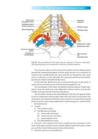
Fig. 213◊The root of the neck. For clarity, only the vagus nerve is shown on the right
and only the phrenic nerve on the left, as this lies on scalenus anterior.
The first part arches over the dome of the pleura and lies deeply placed
beneath the sternocleidomastoid and the strap muscles. It is crossed at its
origin by the carotid sheath and, more laterally, by the phrenic and vagus
nerves. At this site, on the right side, the vagus gives off its recurrent laryn-
geal branch which hooks behind the artery.
On the left side, the thoracic duct crosses the first part of the artery to
open into the commencement of the left branchiocephalic vein.
The second part of the artery lies behind scalenus anterior which sepa-
rates it from the subclavian vein. Behind lie scalenus medius and also the
middle and upper trunks of the brachial plexus.
The third part extends to the lateral border of the first rib against which
it can be compressed and its pulse easily felt, since here it is just below the
deep fascia. Immediately behind the artery is the lower trunk of the brachial
plexus which is, in fact, responsible for the ‘subclavian groove’ on the first rib.
Its branches are:
•◊◊1st part
1◊◊The vertebral artery
2◊◊The thyrocervical trunk:
(a)◊◊inferior thyroid artery
(b)◊◊transverse cervical artery
(c)◊◊suprascapular artery
3◊◊The internal thoracic artery
•◊◊2nd part — the costocervical trunk (supplying deep structures of the
neck via its deep cervical branch, and the superior intercostal artery, which
gives off the 1st and 2nd posterior intercostal arteries).
•◊◊3rd part—gives no constant branch.