
Page 313 - Clinical Anatomy
P. 313
ECA5 7/18/06 6:50 PM Page 298
298 The head and neck
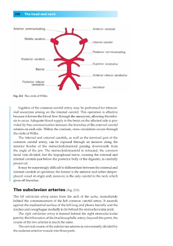
Fig. 212◊The circle of Willis.
Ligation of the common carotid artery may be performed for intracra-
nial aneurysm arising on the internal carotid. This operation is effective
because it lowers the blood flow through the aneurysm, allowing thrombo-
sis to occur. Adequate blood supply to the brain on the affected side is pro-
vided by free communication between the branches of the external carotid
arteries on each side. Within the cranium, cross-circulation occurs through
the circle of Willis.
The internal and external carotids, as well as the terminal part of the
common carotid artery, can be exposed through an incision along the
anterior border of the sternocleidomastoid passing downwards from
the angle of the jaw. The sternocleidomastoid is retracted, the common
facial vein divided, but the hypoglossal nerve, crossing the external and
internal carotids just below the posterior belly of the digastric, is carefully
preserved.
It may be surprisingly difficult to differentiate between the external and
internal carotids at operation; the former is the anterior and rather deeper-
placed vessel at origin and, morever, is the only carotid in the neck which
gives off branches.
The subclavian arteries (Fig. 213)
The left subclavian artery arises from the arch of the aorta, immediately
behind the commencement of the left common carotid artery. It ascends
against the mediastinal surface of the left lung and pleura laterally and the
trachea and oesophagus medially to lie behind the sternoclavicular joint.
The right subclavian artery is formed behind the right sternoclavicular
joint by the bifurcation of the brachiocephalic artery; beyond this point, the
course of the two arteries is much the same.
The cervical course of the subclavian arteries is conveniently divided by
the scalenus anterior muscle into three parts.