
Page 320 - Clinical Anatomy
P. 320
ECA5 7/18/06 6:51 PM Page 305
The veins of the head and neck 305
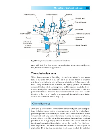
Fig. 217◊The great veins of the neck and their tributaries.
cates with its fellow then passes outwards, deep to the sternocleidomas-
toid, to enter the external jugular vein.
The subclavian vein
This is the continuation of the axillary vein and extends from its commence-
ment at the outer border of the first rib to the medial border of scalenus
anterior, where it joins the internal jugular vein to form the brachiocephalic
vein. During its short course it crosses, and lightly grooves, the superior
surface of the first rib. It arches upwards and then passes medially, down-
wards and slightly forwards to its termination behind the sternoclavicular
joint. On the left side it receives the termination of the thoracic duct. Its only
tributary is the external jugular vein. Anteriorly the vein is related to the
clavicle and subclavius muscle (Fig. 217).
Clinical features
Techniques of central venous catheterization are now of great clinical impor-
tance both to measure central venous pressure (c.v.p.), for practical pur-
poses the pressure within the right atrium, and also to allow rapid blood
replacement and long-term intravenous feeding by means of glucose,
amino acids and fats. The internal jugular vein can be cannulated by direct
puncture in the triangular gap between the sternal and clavicular heads of
the sternocleidomastoid immediately above the clavicle. Feel this land-
mark on yourself. The needle is inserted near the apex of this triangle at an
angle of 30–40° to the skin surface and is advanced caudally towards the