
Page 319 - Clinical Anatomy
P. 319
ECA5 7/18/06 6:51 PM Page 304
304 The head and neck
it continues the sigmoid sinus) to its termination behind the sternal extrem-
ity of the clavicle, where it joins the subclavian vein to form the brachio-
cephalic vein.
It lies lateral first to the internal and then to the common carotid artery
within the carotid sheath and its relations are therefore identical with these
vessels (Fig. 210). The deep cervical chain of lymph nodes lies close against
the vein and, if involved by malignant or inflammatory disease, may
become densely adherent to the vein. Tearing of the jugular vein for this
reason is far from rare in dissections of tuberculous cervical lymph nodes.
Its tributaries are:
1◊◊the pharyngeal venous plexus;
2◊◊the common facial vein;
3◊◊the lingual vein;
4◊◊the superior and middle thyroid veins.
Superficial veins
The arrangement of the superficial veins of the head and neck are some-
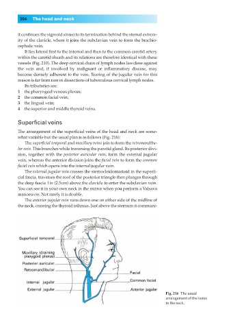
what variable but the usual plan is as follows (Fig. 216):
The superficial temporal and maxillary veins join to form the retromandibu-
lar vein. This branches while traversing the parotid gland. Its posterior divi-
sion, together with the posterior auricular vein, form the external jugular
vein, whereas the anterior division joins the facial vein to form the common
facial vein which opens into the internal jugular vein.
The external jugular vein crosses the sternocleidomastoid in the superfi-
cial fascia, traverses the roof of the posterior triangle then plunges through
the deep fascia 1in (2.5cm) above the clavicle to enter the subclavian vein.
You can see it in your own neck in the mirror when you perform a Valsava
manoeuvre. Not rarely it is double.
The anterior jugular vein runs down one on either side of the midline of
the neck, crossing the thyroid isthmus. Just above the sternum it communi-
Fig. 216◊The usual
arrangement of the veins
in the neck.