
Page 337 - Clinical Anatomy
P. 337
ECA5 7/18/06 6:51 PM Page 322
322 The head and neck
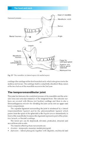
Fig. 227◊The mandible: (a) lateral aspect, (b) medial aspect.
cartilage (the cartilage of the first branchial arch, which also gives rise to the
malleus and incus). The cartilage itself is completely absorbed. Bony union
of the two halves of the mandible occurs in the 2nd year.
The temporomandibular joint
This joint lies between the condyloid process of the mandible and the artic-
ular fossa and articular eminence of the temporal bone. The articular sur-
faces are covered with fibrous (not hyaline) cartilage and there is also a
fibrocartilaginous articular disc dividing the joint cavity into an upper and
lower compartment.
The capsular ligament surrounding the joint is reinforced by a lateral
temporomandibular ligament and by the sphenomandibular ligament which
passes from the spine of the sphenoid to the lingular process immediately in
front of the mandibular foramen; this ligament represents part of the primi-
tive 1st arch, or Meckel’s cartilage.
The lower jaw can be depressed, elevated, protruded, retracted and
moved from side to side.
The muscles effecting these movements are:
•◊◊elevation—temporalis, masseter, medial pterygoid;
•◊◊depression — lateral pterygoid, together with digastric, mylohyoid and