
Page 333 - Clinical Anatomy
P. 333
ECA5 7/18/06 6:51 PM Page 318
318 The head and neck
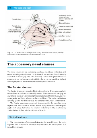
Fig. 225◊The lateral wall of the right nasal cavity; the conchae have been partially
removed to show structures which drain into the nose.
The accessory nasal sinuses
The nasal sinuses are air-containing sacs lined by ciliated epithelium and
communicating with the nasal cavity through narrow, and therefore easily
occluded, channels (Fig. 225). The maxillary antrum and sphenoid sinuses
are present in a rudimentary state at birth, the rest become evident at about
the 8th year, but all become fully formed only in adolescence.
The frontal sinuses
The frontal sinuses are contained in the frontal bone. They vary greatly in
size and one or both are occasionally absent. In section each is roughly tri-
angular, its anterior wall forming the prominence of the forehead, its pos-
terosuperior wall lying adjacent to the frontal lobe of the brain, and its floor
abutting against the ethmoid cells, the roof of the nasal fossa and the orbit.
The frontal sinuses are separated from each other by a median bony
septum, and each in turn is further broken up by a number of incomplete
septa. Each sinus drains into the anterior part of the middle nasal meatus
via the infundibulum into the hiatus semilunaris.
Clinical features
1◊◊The close relation of the frontal sinus to the frontal lobe of the brain
explains how infection of this sinus may result in the development of a
frontal lobe abscess.