
Page 56 - Clinical Anatomy
P. 56
ECA1 7/18/06 6:31 PM Page 41
The mediastinum 41
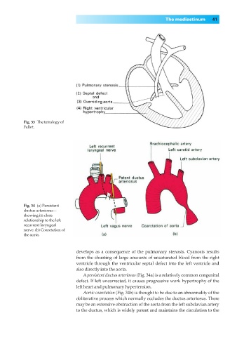
Fig. 33◊The tetralogy of
Fallot.
Fig. 34◊(a) Persistent
ductus arteriosus—
showing its close
relationship to the left
recurrent laryngeal
nerve. (b) Coarctation of
the aorta.
develops as a consequence of the pulmonary stenosis. Cyanosis results
from the shunting of large amounts of unsaturated blood from the right
ventricle through the ventricular septal defect into the left ventricle and
also directly into the aorta.
Apersistent ductus arteriosus (Fig. 34a) is a relatively common congenital
defect. If left uncorrected, it causes progressive work hypertrophy of the
left heart and pulmonary hypertension.
Aortic coarctation (Fig. 34b) is thought to be due to an abnormality of the
obliterative process which normally occludes the ductus arteriosus. There
may be an extensive obstruction of the aorta from the left subclavian artery
to the ductus, which is widely patent and maintains the circulation to the