Page 54 - Clinical Anatomy
P. 54

ECA1  7/18/06  6:31 PM  Page 39


                                                                            The mediastinum     39


                                                                                     Left common
                                                                                     carotid artery
                          Brachiocephalic                                            Left subclavian
                                  artery                                             artery
                          Right pulmonary
                                  artery                                             Ductus arteriosus
                                  Aorta
                                                                                     Left pulmonary
                                Superior                                             artery
                               vena cava
                               Septum II
                                                                                     Pulmonary trunk
                           Foramen ovale
                                Septum I


                                                                                     Aorta


                                 Inferior
                               vena cava


                                                                                     Umbilical arteries

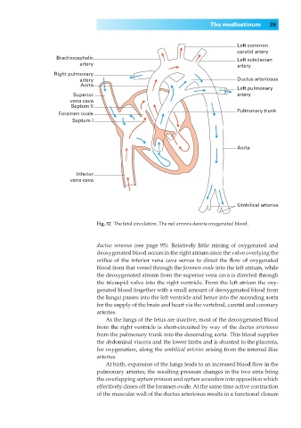
                                        Fig. 32◊The fetal circulation. The red arrows denote oxygenated blood.


                                        ductus venosus (see page 95). Relatively little mixing of oxygenated and
                                        deoxygenated blood occurs in the right atrium since the valve overlying the
                                        orifice of the inferior vena cava serves to direct the flow of oxygenated
                                        blood from that vessel through the foramen ovale into the left atrium, while
                                        the deoxygenated stream from the superior vena cava is directed through
                                        the tricuspid valve into the right ventricle. From the left atrium the oxy-
                                        genated blood (together with a small amount of deoxygenated blood from
                                        the lungs) passes into the left ventricle and hence into the ascending aorta
                                        for the supply of the brain and heart via the vertebral, carotid and coronary
                                        arteries.
                                          As the lungs of the fetus are inactive, most of the deoxygenated blood
                                        from the right ventricle is short-circuited by way of the ductus arteriosus
                                        from the pulmonary trunk into the descending aorta. This blood supplies
                                        the abdominal viscera and the lower limbs and is shunted to the placenta,
                                        for oxygenation, along the umbilical arteries arising from the internal iliac
                                        arteries.
                                          At birth, expansion of the lungs leads to an increased blood flow in the
                                        pulmonary arteries; the resulting pressure changes in the two atria bring
                                        the overlapping septum primum and septum secundum into apposition which
                                        effectively closes off the foramen ovale. At the same time active contraction
                                        of the muscular wall of the ductus arteriosus results in a functional closure
   49   50   51   52   53   54   55   56   57   58   59