
Page 53 - Clinical Anatomy
P. 53
ECA1 7/18/06 6:31 PM Page 38
38 The Thorax
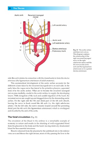
Fig. 31◊The aortic arches
and their derivatives.
This diagram explains
the relationship of the
right recurrent laryngeal
nerve to the right
subclavian artery and the
left nerve to the aortic
arch and the ligamentum
arteriosum (or to a patent
ductus arteriosus).
side this arch retains its connection with the dorsal aorta to form the ductus
arteriosus (the ligamentum arteriosum of adult anatomy).
This asymmetrical development of the aortic arches accounts for the
different course taken by the recurrent laryngeal nerve on each side. In the
early fetus the vagus nerve lies lateral to the primitive pharynx, separated
from it by the aortic arches. What are to become the recurrent laryngeal
nerves pass medially, caudal to the aortic arches, to supply the developing
larynx. With elongation of the neck and caudal migration of the heart, the
recurrent nerves are caught up and dragged down by the descending aortic
arches. On the right side the 5th and distal part of the 6th arch absorb,
leaving the nerve to hook round the 4th arch (i.e. the right subclavian
artery). On the left side, the nerve remains looped around the persisting
distal part the 6th arch (the ligamentum arteriosum) which is overlapped
and dwarfed by the arch of the aorta.
The fetal circulation (Fig. 32)
The circulation of the blood in the embryo is a remarkable example of
economy in nature and results in the shunting of well-oxygenated blood
from the placenta to the brain and the heart, leaving relatively desaturated
blood for less essential structures.
Blood is returned from the placenta by the umbilical vein to the inferior
vena cava and thence the right atrium, most of it by-passing the liver in the