
Page 154 - Critical Care Notes
P. 154
4223_Tab05_141-174 29/08/14 8:28 AM Page 148
NEURO
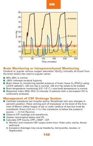
90
80
A-waves
70
60
mm Hg 50 B-waves
40
30
20 C
10 N
0
0 60 120 180 240 300
Time (minutes)
Brain Monitoring or Intraparenchymal Monitoring
Cerebral or jugular venous oxygen saturation (SjvO 2 ) (virtually all blood from
the brain drains into internal jugular veins):
■ 60%–80% is normal.
■ <50% indicates cerebral hypoxia.
■ Brain tissue O 2 monitoring (partial pressure of brain tissue O 2 [PbtO 2 ] using
LICOX catheter): >25 mm Hg is normal; <20 mm Hg needs to be treated.
■ Brain temperature monitoring: 0.5°–1.0° C > core body temperature is normal.
■ Bispectral index (BIS): EEG of critically ill patients with a decreased LOC is
continually analyzed.
Management of CSF Drainage System
■ Calibrate transducer per hospital policy. Recalibrate with any changes in
patient’s position. Place venting port of transducer at the level of the fora-
men of Monro (either tragus of ear or outer canthus of eye but must be
consistent). Every 2.54 cm (1 in.) the transducer is below the pressure
source → –2 mm Hg error.
■ Observe ICP readings and waveforms.
■ Assess neurological status and VS.
■ Calculate CPP hourly; CPP = MAP – ICP.
■ Monitor and measure CSF output every hour. Note color, clarity, blood,
sediment.
■ Excessive drainage may cause headache, tachycardia, nausea, or
diaphoresis.
148