
Page 273 - Cardiac Nursing
P. 273
LWBK340-c11_p245-266.qxd 6/29/09 10:21 PM Page 249 Aptara Inc.
C HAP TE R 11 / Laboratory Tests Using Blood 249
Myocardial Proteins
BIOCHEMICAL MARKERS OF
MYOCARDIAL INJURY Troponins
Troponins are protein complexes that regulate the calcium-depend-
ent interaction of myosin with actin in the muscle contractile ap-
The internal environment of the healthy person is in a state of bal- paratus of striated muscle. They are found in both cardiac and skele-
ance with respect to water, electrolytes, energy storage and use, tal muscle. Three isotypes have been identified: troponin-I (cTnI),
and metabolic end products. Stability is maintained through troponin-T (cTnT), and troponin-C (cTnC). Troponins T and I
homeostatic mechanisms that regulate the activities of cells and are both found in the myocardium. Troponin-T binds the troponin
organs. During periods of critical illness, a disruption in cell complex to tropomyosin, and troponin-I inhibits the muscle con-
membrane stability may cause chemical substances that are re- traction in the absence of calcium and troponin-C. Troponin-C
2
sponsible for intracellular homeostatic mechanisms to appear in lacks cardiac specificity; therefore, it is the least studied of the tro-
the blood. Frequent evaluation of blood results is a means by ponins and has no assay available in the clinical setting.
which the status of the internal environment and the extent and Because of the high specificity and sensitivity for detecting my-
nature of tissue damage can be monitored. These blood tests can ocardial injury, troponin has become the most important addition
be run expeditiously and require small sample volumes, and pro- to clinical laboratory testing for assessment of myocardial injury. 12
vide important information concerning the diagnosis and man- In patients with acute coronary syndrome, troponin is an enor-
agement of patients. 19 mously useful biochemical marker in the early diagnosis of MI be-
Certain intracellular enzymes and proteins are rarely found in cause it is either low or undetectable in healthy people, but in the
measurable amounts in the blood of healthy people. However, af- event of an MI, is detectable as early as 2 to 3 hours after injury. 19
ter an event leading to cellular injury or death, these substances Testing for troponin is typically done at the time of the initial
may leak into the blood. A continued question with ongoing re- workup for suspected acute coronary syndrome or myocardial
search is the extent to which reversible cell damage can cause pro- damage and then 6 to 9 hours later. An additional sample may be
tein leakage. 20 Because of the importance of the timing of the ap- measured between 12 and 24 hours if biochemical markers have
pearance (and disappearance) of enzymes and proteins in the not shown elevation and MI is still suspected. 22,23 Because most
blood, it is crucial that ordered tests are drawn on time. It is troponin is so tightly bound to muscle, it is released slowly and
equally important that the date and time of the blood draw are may remain detectable for 1 to 2 weeks post-MI. This late-phase
22
noted on the laboratory slip so that the temporal sequence of the presence of troponin represents death of the contractile apparatus.
rise and fall can be established by those interpreting the results. Because troponin remains elevated longer than CK-MB and is
Over the years as more specific biochemical markers of my- more specific than LDH, troponin is now the preferred test for
ocardial injury have become available, detecting MI has become patients who seek medical attention more than 24 to 48 hours af-
more accurate. The original marker, glutamine-oxaloacetic ter myocardial injury. The appearance of troponin in the blood in-
transaminase was replaced by lactate dehydrogenase (LDH) and dicates necrosis or injury to the myocardium and follows a pre-
later by CK and CK-MB. Troponin has now become the preferred dictable rise and fall over a specified time. See Table 11-2 and
laboratory test for diagnosing MI and the other markers are be- Figure 11-1 for the typical appearance, peak, and disappearance of
coming obsolete. 21 Initial diagnosis of MI, reinfarction, or other various biochemical markers and enzymes.
types of myocardial damage is made through evaluation of clini- In patients with ST-segment elevation MI, percutaneous coro-
cal signs and symptoms, 12-lead ECG, biochemical markers in- nary intervention (PCI) or fibrinolytic therapy should not be de-
cluding myocardial proteins (troponins) and if troponin not avail- layed waiting for biochemical marker evaluation. 21 For other pa-
able, cardiac enzymes (see Chapter 22). 22 A comparison of the tients with suspected cardiac symptoms, troponin is used along
sensitivity and specificity of various tests to detect myocardial in- with clinical signs and symptoms and 12-lead ECG to make
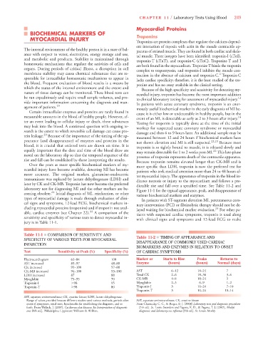
jury is in Table 11-1.
Table 11-1 ■ COMPARISON OF SENSITIVITY AND Table 11-2 ■ TIMING OF APPEARANCE AND
SPECIFICITY OF VARIOUS TESTS FOR MYOCARDIAL DISAPPEARANCE OF COMMONLY USED CARDIAC
INFARCTION
BIOMARKERS AND ENZYMES IN RELATION TO ONSET
Test Sensitivity at Peak (%) Specificity (%) OF CARDIAC SYMPTOMS
Electrocardiogram 63–84 100 Marker or Starts to Rise Peaks Returns to
AST increased 89–97 48–88 Enzyme (hours) (hours) Normal (days)
CK increased 93–100 57–88
CK-MB increased 94–100 93–100 AST 6–12 18–24 7
LDH increased 87 88 Total CK 2–6 18–36 3–6
Myoglobin 75–95 70 CK-MB 4–8 18–24 3
Troponin-I 98 95 Myoglobin 2–3 6–9 1–2
Troponin-T 98 80 Troponin-I 3 10–24 7–10
Troponin-T 3 10–24 10–14
AST, aspartate aminotransferase; CK, creatine kinase; LDH, lactate dehydrogenase.
Range of values provided because different studies used various methods, periods after AST, aspartate aminotransferase; CK, creatine kinase
onset of symptoms, serial tests, benchmarks for establishing the diagnosis, and so From Chernecky, C. C., & Berger, B. J. [2008] Laboratory tests and diagnostic procedures
forth. From Wallach, J. [2007]. Cardiovascular diseases. In Interpretation of diagnostic [5th ed.]. St. Louis: Saunders and Pagana, K. D., & Pagana, T. J. [2007], Mosby’s
tests [8th ed.]. Philadelphia: Lippincott Williams & Wilkins. diagnostic and laboratory test reference [8th ed.]. St. Louis: Mosby.