
Page 118 - untitled
P. 118
AAAC51 21/5/05 10:58 AM Page 117
Ligaments of the foot
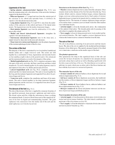
• Muscles: extensor digitorum brevis arises from the calcaneus. Other
• Spring (plantar calcaneonavicular) ligament (Fig. 51.2): runs
muscles insert on the dorsum of the foot but arise from the leg. These
from the sustentaculum tali to the tuberosity of the navicular forming a
include: tibialis anterior, extensor hallucis longus, extensor digitorum
support for the head of the talus. Structures on the dorsum of the foot (Fig. 51.1)
• Bifurcate ligament: is Y shaped and runs from the anterior part of longus, peroneus tertius and peroneus brevis. Each tendon of extensor
the calcaneus to the cuboid and navicular bones. It reinforces the digitorum longus is joined on its lateral side by a tendon from extensor
capsule of the talocalcaneonavicular joint. digitorum brevis. The tendons of extensor digitorum longus and per-
• Long plantar ligament (Figs 51.2 and 50.3): runs from the under- oneus tertius share a common synovial sheath whilst the other tendons
surface of the calcaneus to the cuboid and bases of the lateral meta- have individual sheaths.
tarsals. The ligament runs over the tendon of peroneus longus. • Arterial supply: is from the dorsalis pedis arteryathe continuation
• Short plantar ligament: runs from the undersurface of the calca- of the anterior tibial artery. The dorsalis pedis ends by passing to the
neus to the cuboid. sole where it completes the plantar arch (p. 95).
• Medial and lateral (talocalcaneal) ligaments: strengthen the • Nerve supply: is from the deep peroneal nerve via its medial and lat-
capsule of the talocalcaneal joint. eral terminal branches. The latter supplies extensor digitorum brevis
• Interosseous talocalcaneal ligament: runs in the sinus tarsi, a whereas the former receives cutaneous branches from the skin.
tunnel formed by deep sulci on the talus and calcaneus.
• Deep transverse metacarpal ligaments: join the plantar ligaments The sole of the foot
of the metatarsophalangeal joints of the five toes. The sole is described as consisting of an aponeurosis and four muscle
layers. The skin of the sole is supplied by the medial and lateral plantar
The arches of the foot branches of the tibial nerve. The medial calcaneal branch of the tibial
The integrity of the foot is maintained by two longitudinal (medial and nerve innervates a small area on the medial aspect of the heel.
lateral) arches and a single transverse arch. The arches are held
together by a combination of bony, ligamentous and muscular factors The plantar aponeurosis
so that standing weight is taken on the posterior part of the calcaneum This aponeurosis lies deep to the superficial fascia of the sole and
and the metatarsal heads as a result of the integrity of the arches. covers the 1st layer of muscles. It is attached to the calcaneus behind
• Medial longitudinal arch (see Fig. 50.2): comprises calcaneus, talus and sends a deep slip to each toe as well as blending superficially with
(the apex of the arch), navicular, the three cuneiforms and three medial the skin creases at the base of the toes. The slips that are sent to each toe
metatarsals. The arch is bound together by the spring ligament, muscles split into two parts which pass on either side of the flexor tendons and
and supported from above by tibialis anterior and posterior. fuse with the deep transverse metatarsal ligaments.
• Lateral longitudinal arch (see Fig. 50.2): comprises calcaneus,
cuboid and the two lateral metatarsals. The arch is bound together by The muscular layers of the sole
the long and short plantar ligaments and supported from above by per- • 1st layer consists of: abductor hallucis, flexor digitorum brevis and
oneus longus and brevis. abductor digiti minimi (Fig. 51.3).
• Transverse arch: comprises the cuneiforms and bases of the meta- • 2nd layer consists of: flexor digitorum accessorius, the lumbricals
tarsals. The arch is bound together by the deep transverse ligament, and the tendons of flexor digitorum longus and flexor hallucis longus
plantar ligaments and the interossei. It is supported from above by per- (Fig. 51.4).
oneus longus and brevis. • 3rd layer consists of: flexor hallucis brevis, adductor hallucis and
flexor digiti minimi brevis (Fig. 51.5).
The dorsum of the foot (Fig. 51.1) • 4th layer consists of: the dorsal and plantar interossei and the ten-
The skin of the dorsum of the foot is supplied by cutaneous branches of dons of peroneus longus and tibialis posterior.
the superficial peroneal, deep peroneal, saphenous and sural nerves.
The dorsal venous arch lies within the subcutaneous tissue overlying Neurovascular structures of the sole
the metatarsal heads. It receives blood from most of the superficial tis- • Arterial supply: is from the posterior tibial artery which divides into
sues of the foot via digital and communicating branches. The great medial and lateral plantar branches. The latter branch contributes the
saphenous vein commences from the medial end of the arch and the major part of the deep plantar arch (p. 95).
small saphenous vein from the lateral end. • Nerve supply: is from the tibial nerve which also divides into medial
and lateral plantar branches (p. 101).
The ankle and foot II 117