
Page 119 - untitled
P. 119
AAAC52 21/5/05 10:58 AM Page 118
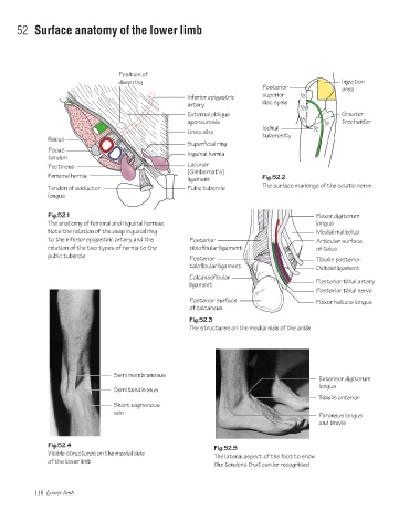
52 Surface anatomy of the lower limb
Position of
deep ring Injection
Posterior area
Inferior epigastric superior 1 /3
artery iliac spine 1 /3
External oblique Greater
1 /3
aponeurosis 1 /2 trochanter
Ischial 1 /2
Linea alba tuberosity
Iliacus
Superficial ring
Psoas Inguinal hernia
tendon
Lacunar
Pectineus
(Gimbernat's)
Femoral hernia Fig.52.2
ligament
The surface markings of the sciatic nerve
Tendon of adductor Pubic tubercle
longus
Fig.52.1 Flexor digitorum
The anatomy of femoral and inguinal herniae. longus
Note the relation of the deep inguinal ring Medial malleolus
to the inferior epigastric artery and the Posterior Articular surface
relation of the two types of hernia to the tibiofibular ligament of talus
pubic tubercle
Posterior Tibialis posterior
talofibular ligament Deltoid ligament
Calcaneofibular
Posterior tibial artery
ligament
Posterior tibial nerve
Posterior surface Flexor hallucis longus
of calcaneus
Fig.52.3
The structures on the medial side of the ankle
Semi membranosus
Extensor digitorum
longus
Semi tendinosus
Tibialis anterior
Short saphenous
vein
Peroneus longus
and brevis
Fig.52.4
Fig.52.5
Visible structures on the medial side
The lateral aspect of the foot to show
of the lower limb
the tendons that can be recognised
118 Lower limb