Page 127 - untitled
P. 127

AAAC56  21/5/05  10:56 AM  Page 126
           56 Spinal nerves and cranial nerves I–IV
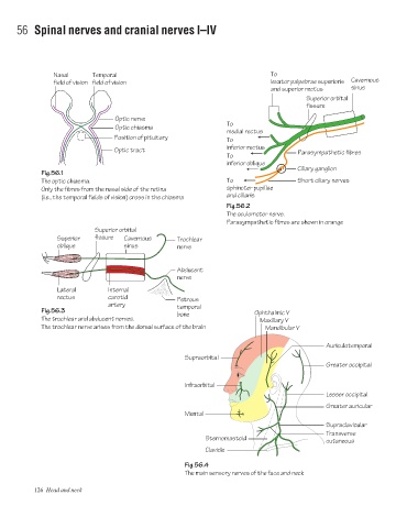
                      Nasal      Temporal                                             To
                      field of vision field of vision                                 levator palpebrae superioris  Cavernous
                                                                                      and superior rectus     sinus
                                                                                                 Superior orbital
                                                                                                 fissure
                                        Optic nerve
                                                                         To
                                        Optic chiasma
                                                                         medial rectus
                                        Position of pituitary
                                                                         To
                                                                         inferior rectus
                                        Optic tract                                           Parasympathetic fibres
                                                                         To
                                                                         inferior oblique
                                                                                              Ciliary ganglion
                  Fig.56.1
                  The optic chiasma.                                     To                   Short ciliary nerves
                  Only the fibres from the nasal side of the retina      sphincter pupillae
                  (i.e., the temporal fields of vision) cross in the chiasma   and ciliaris
                                                                         Fig.56.2
                                                                         The oculomotor nerve.
                                                                         Parasympathetic fibres are shown in orange
                                  Superior orbital
                       Superior   fissure  Cavernous      Trochlear
                       oblique            sinus           nerve


                                                          Abducent
                                                          nerve
                       Lateral        Internal
                       rectus         carotid             Petrous
                                      artery              temporal
                  Fig.56.3
                                                          bone                   Ophthalmic V
                  The trochlear and abducent nerves.                               Maxillary V
                  The trochlear nerve arises from the dorsal surface of the brain   Mandibular V

                                                                                                       Auriculotemporal

                                                             Supraorbital
                                                                                                      Greater occipital

                                                             Infraorbital
                                                                                                      Lesser occipital
                                                                                                      Greater auricular
                                                             Mental
                                                                                                      Supraclavicular
                                                                                                      Transverse
                                                                   Sternomastoid
                                                                                                      cutaneous
                                                                   Clavicle
                                                             Fig.56.4
                                                             The main sensory nerves of the face and neck

                126 Head and neck
   122   123   124   125   126   127   128   129   130   131   132