
Page 137 - untitled
P. 137
AAAC61 21/5/05 11:03 AM Page 136
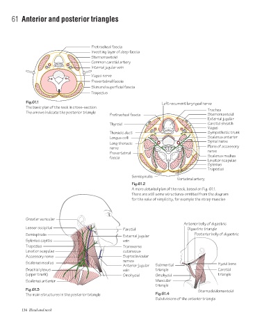
61 Anterior and posterior triangles
Pretracheal fascia
Investing layer of deep fascia
Sternomastoid
Common carotid artery
Internal jugular vein
Vagus nerve
Prevertebral fascia
Skin and superficial fascia
Trapezius
Fig.61.1 Left recurrent laryngeal nerve
The basic plan of the neck in cross-section.
Trachea
The arrows indicate the posterior triangle
Pretracheal fascia Sternomastoid
External jugular
Thyroid Carotid sheath
Vagus
Thoracic duct Sympathetic trunk
Longus colli Scalenus anterior
Spinal nerve
Long thoracic
Plane of accessory
nerve
nerve
Prevertebral
fascia Scalenus medius
Levator scapulae
Splenius
Trapezius
Semispinalis
Vertebral artery
Fig.61.2
A more detailed plan of the neck, based on Fig. 61.1.
There are still some structures omitted from the diagram
for the sake of simplicity, for example the strap muscles
Greater auricular
Anterior belly of digastric
Lesser occipital
Parotid Digastric triangle
Semispinalis External jugular Posterior belly of digastric
Splenius capitis vein
Trapezius Transverse
Levator scapulae cutaneous
Accessory nerve Supraclavicular
nerves
Scalenus medius Hyoid bone
Anterior jugular Submental
Brachial plexus vein triangle Carotid
(upper trunk) Omohyoid Omohyoid triangle
Scalenus anterior Muscular
triangle
Fig.61.3 Sternocleidomastoid
The main structures in the posterior triangle Fig.61.4
Subdivisions of the anterior triangle
136 Head and neck