
Page 229 - Color Atlas Of Pathophysiology (S Silbernagl Et Al, Thieme 2000)
P. 229
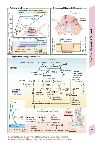
A.  Coronary Reserve             B.  Collateral Myocardial Perfusion
               Resistance in distal coronary aa.:     Left
               normal    50%            Epi-          coronary artery
                           1/4 (vasodilation)
        Poststenotic pressure, P ps  (mmHg)  80  80% 75%  70%  (%) closed  (after van der Werf)  cardium  Endocardium  circulation
        100
                           Normal
                                                             Collateral
         60
         40
                   P Ao = 90
                            P ps
         20
                     Proximal stenosis
                  100
                               250
                           200 .
                       150
              50
                                   –1
                             Q (mL · min )
        Diameter decreased  0 (% closed)  80% closed 70% closed Regulatory Normal  Myocardial circulation  (mL · min –1  · g –1 )  Subendocardial  1.0 (afterh Kirk and Jennings)  Myocardial Infarction I
                                                Subepicardial
                             50% closed
                 75% closed
                                .
                                                               0.2
                           range of Q
                                                               0
                           (distal vasodilation)
        C.  Myocardial Energy Metabolism        Ischemic zone          Plate 7.23
                                     Little ATP
               Glucose  G-6-P     Pyruvate  Acetyl-CoA
                                                  O 2
                                   NADH
         Normal                          FADH 2
                                NAD +               ATP  Normal
                         Lactate        FAD              myocardial
                         (consumption) Free
                                  fatty acids   CO 2     contraction
                                                         Creatine
                                     Little ATP         phosphate
               Glucose  G-6-P     Pyruvate  Acetyl-CoA     store
                  Glycogen         NADH          O 2
                     store                                    Cr∼ P
          Ischemic                 NAD +           Little  Little ATP
                                                   ATP
          anoxia           +  Lactate  Free  FAD
          (early stage)   H  (formation)  fatty acids  CO 2  ATP deficiency  Cr
                                        Intracellular
                                        fat deposition
                                                       Abnormal
            CK           Reversible cell damage        myocardial
                                                       contraction
             SGOT
                                      +
                             No removal of H  and Lactate  Angina
                                                  pectoris
                     Ischemia  Acidosis  Lactate
                LDH 1
                     (longer than       accumulation
                     15–20 min)  Inhibits glycolysis (and others)
                                        ATP deficiency
        Normal range    Enzyme
        0 2 4 6 8 10 12  released  Irreversible cell damage  Infarction  219
                        into plasma
                 Days
       Silbernagl/Lang, Color Atlas of Pathophysiology © 2000 Thieme
       All rights reserved. Usage subject to terms and conditions of license.