
Page 232 - Color Atlas Of Pathophysiology (S Silbernagl Et Al, Thieme 2000)
P. 232
"
(tissue plasminogen activator [t-PA], anti- If sizeable portions of the myocardium die,
thrombin III, heparin sulfate, protein C, enzymes are released from the myocardial
thrombomodulin, and prostacyclin). cells into the bloodstream. It is not so much
Rare causes of MI are inflammatory vascular the level of enzyme concentrations as the tem-
diseases, embolism (endocarditis; valve pros- poral course of their maxima that is important
thesis), severe coronary spasm (e.g., after tak- in the diagnosis of MI. Myocardial creatine ki-
ing cocaine), increased blood viscosity as well nase (CK-MB [MB = muscle, brain]) reaches its
as a markedly raised O 2 demand at rest (e.g., peak on day 1, aspartate aminotransferase
in aortic stenosis). (ASAT) on day 2, and myocardial lactate dehy-
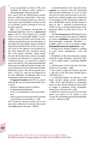
ECG (→ F). A prominent characteristic of drogenase (LDH 1 ) on days three to five (→ C,
transmural infarction (tmI) is an abnormal Q bottom).
wave (→ F1) of > 0.04 seconds and a voltage Possible consequences of MI depend on site,
that is > 25% of overall QRS voltage. It occurs extent, and scarring of the infarct. In addition
to various arrhythmias, among them acutely
within one day and is due to the necrotic myo-
Heart and Circulation that when this myocardial segment should be p.186ff.), there is a risk of a number of mor-
cardium not providing any electrical signal, so
life-threatening ventricular fibrillation (→
phological/mechanical complications (→ G):
depolarized (within the first 0.04 s), the excita-
! Tearing of the chordae tendineae resulting
tion vector of the opposite, normal portion of
in acute mitral regurgitation (→ G1 and
the heart dominates the summated vector.
p.196);
This “0.04 vector” therefore “points away”
in anterior-wall infarction, it is registered par-
with left-to-right shunting (→ G2 and p. 204);
! Fall in cardiac output (→ G,a) that, together
ticularly in leads V 5 , V 6 , I, and aVL as a large Q
7 from the site of infarction so that, for example, ! Perforation of the interventricular septum
wave (and small R). (In a transmural infarction with
of the posterior wall such Q wave changes can- ! stiffened parts of the ventricular wall (aki-
not be registered with the conventional leads). nesia) due to scarring (→ G,b),
Abnormal Q waves will still be present years ! will result in a high end-diastolic pressure
later (→ F2,3), i.e., they are not diagnostic of (→ G3 and p. 224). Still more harmful than a
an acute infarction. An infarction that is not stiff infarct scar is
transmural usually causes no Q changes. ! a stretchable infarct area, because it will
ST segment elevation in the ECG is a sign of bulge outward during systole (dyskinesia;
ischemic but not (yet) dead myocardial tissue. → G4), which will therefore—at comparably
It occurs large scar area—be more likely to reduce cardi-
– during an anginal attack (see above) ac output to dangerous levels (cardiogenic
– in nontransmural infarction shock) than a stiff scar will (→ G5);
– at the very beginning of transmural infarc- ! Finally, the ventricular wall at the site of the
tion infarct can rupture to the outside so that
– at the margin of a transmural infarction that acutely life-threatening pericardial tamponade
occurred hours to days before (→ F4) occurs (→ G6 and p. 228).
The ST segment returns to normal one to two
days after an MI, but for the next few weeks
the T wave will be inverted (→ F5,F2).
222
Silbernagl/Lang, Color Atlas of Pathophysiology © 2000 Thieme
All rights reserved. Usage subject to terms and conditions of license.