
Page 81 - Color Atlas Of Pathophysiology (S Silbernagl Et Al, Thieme 2000)
P. 81
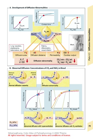
A.  Development of Diffusion Abnormalities


           [O 2 ] blood       P O 2  capillaries  [O 2 ] blood  O 2  ven.


                 O 2  art.       Contact time
               P O 2  cap.                             P O 2  cap.
                                   P O 2  alv.
                         [O 2 ]   art.      [O 2 ] ven.                Abnormalities

                                   P O 2  cap.
                                                       Work
         Lung resection,         Edema,                                Diffusion
         emphysema,              inflammation,
         tuberculosis, etc.      vessel dilation
                    F      d                 K    CO
             Diffusion area  Diffusion distance  Permeability  Cardiac output  Plate 4.3
                 K· F      Diffusion abnormality  [O 2 ] ven.– [O 2 ] art.
                d· CO                         P O2  cap.– P O2  alv.
        B.  Abnormal Diffusion: Concentrations of CO 2  and HbO 2  in Blood

        Venous           Arterial
        blood    5        blood            5
         7  6     13    13  5     4  6.5    13    6 5.5

        1                         2
        Normal diffusion capacity  Diffusion abnormality


                    [CO 2 ] blood       [HbO 2 ] blood


                             Normal                   Normal

                        P CO 2  blood                  P O 2  blood
                             3                       5
                    5  6       17    10 4     7 6.5    30   24 5.5

         P O 2  (kPa)
                    3                       4
         P CO 2  (kPa)  Abnormal diffusion with hyperventilation  Abnormal diffusion with O 2  ventilation  71

       Silbernagl/Lang, Color Atlas of Pathophysiology © 2000 Thieme
       All rights reserved. Usage subject to terms and conditions of license.