
Page 531 - ACCCN's Critical Care Nursing
P. 531
508 P R I N C I P L E S A N D P R A C T I C E O F C R I T I C A L C A R E
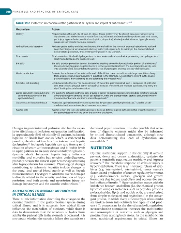
TABLE 19.2 Protective mechanisms of the gastrointestinal system and impact of critical illness 1,3-12
Mechanism Action
Motility Propels bacteria through the GI tract. In critical illness, motility may be altered because of enteric nerve
impairment and altered smooth muscle function, inflammation (mediated by cytokines and nitric oxide),
gut injury, hypoperfusion, medications (opioids, dopamine), electrolyte disturbances, hyperglycaemia,
sepsis and increased intracranial pressure. 3
Hydrochloric acid secretion Reduces gastric acidity and destroys bacteria. Parietal cells in the stomach produce hydrochloric acid and
keep the intragastric environment relatively acidic (pH approx 4.0). An acidic pH has bactericidal and
bacteriostatic properties, thus limiting overgrowth in the stomach.
4
Bicarbonate Bicarbonate ions bind with hydrogen ions to form water and carbon dioxide, preventing the hydrogen ions
(acid) from damaging the duodenal wall. 5
Bile salts Bile salts provide protection against bacteria by breaking down the liposaccharide portion of endotoxins, 6
thereby detoxifying gram-negative bacteria in the gastrointestinal tract. The deconjugation of bile salts
into secondary bile acids inhibits the proliferation of pathogens and may destroy their cell walls. 7
Mucin production Prevents the adhesion of bacteria to the wall of the GI tract. Mucous cells secrete large quantities of very
thick, alkaline mucus (approximately 1 mm thick in the stomach). Glycoproteins present in the mucus
prevent bacteria from adhering to and colonising the mucosal wall. 8
Epithelial cell shedding Limits bacterial adhesion. The mucosal lining of the entire gastrointestinal tract is composed of epithelial
cells that create a physical barrier to bacterial invasion. These cells are replaced approximately every 3–5
days limiting bacterial colonisation.
9
Zonea occludulns (tight junctions The junctions between epithelial cells provide a barrier to microorganisms. Intermediate junctions (zonula
surrounding each cell in the adherens) function primarily in cell–cell adhesion, while the tight junctions (zonula occludens) limit the
epithelial sheet) movement of bacteria and toxins across the gut wall. 10
Gut-associated lymphoid tissue Protection against bacterial invasion is provided by gut-associated lymphoid tissue, capable of cell-
11
mediated and humoral-mediated immune responses. 12
Kupffer cells Kupffer cells in the liver and spleen provide a back-up defence against pathogens that cross the barrier of
the gastrointestinal wall and enter the systemic circulation. 1
Changes in gastrointestinal perfusion also has the capac- decreased pepsin secretion. It is also possible that secre-
ity to affect hepatic perfusion, oxygenation and function. tion of digestive enzymes might also be influenced
In approximately 50% of critically ill patients, ischaemic by critical illness-induced pancreatitis, although clear
hepatitis or ‘shock liver’ occurs, which is evidenced by data demonstrating this level of dysfunction are
jaundice, elevation of liver function tests or overt hepatic unavailable. 16
23
dysfunction. Ischaemic hepatitis can vary from a mild
elevation of serum aminotransferase and bilirubin levels NUTRITION
in septic patients, to an acute elevation following haemo-
dynamic shock. Ischaemic hepatic injury influences Optimal nutritional support in the critically ill aims to
morbidity and mortality but remains underdiagnosed, prevent, detect and correct malnutrition, optimise the
probably because the clinical signs become apparent long patient’s metabolic state, reduce morbidity and improve
24
after hypoperfusion has occurred. Physiological changes recovery. The metabolic response of stress or injury is
contributing to ischaemic hepatitis include changes to hypermetabolism. There is an increased release of cyto-
the portal and arterial blood supply as well as hepatic kines (e.g. interleukin-1, interleukin-6, tumor necrosis
microcirculation. The degree to which the liver is damaged factor-α) and production of counter-regulatory hormones
is directly related to the severity and duration of hypo- (e.g. catecholamines, cortisol, glucagon and growth
perfusion, and both anoxic and reperfusion injury can hormone) that induce catabolism and oppose the ana-
25
damage hepatocytes and the vascular endothelium. 23 bolic effects of insulin. Hypercatabolism occurs with the
imbalance between anabolism (i.e. the chemical process
ALTERATIONS TO NORMAL METABOLISM by which complex molecules, such as peptides, proteins,
polysaccharides, lipids and nucleic acids, are synthesised
IN CRITICAL ILLNESS from simpler molecules) and catabolism (i.e. the conver-
There is little information describing the changes to the gent process, in which many different types of molecules
exocrine function in the gastrointestinal system during are broken down into relatively few types of end prod-
critical illness, and it is uncertain how critical illness ucts). To compensate for the altered metabolic regulation,
influences the metabolism of nutrients. While there is neuroendocrine stimulation increases the mobilisation
data to demonstrate that the secretion of hydrochloric and consumption of nutrients, such as glycogen and
acid by the parietal cells in the stomach is decreased, it is protein, from existing body stores. As the metabolic rate
not certain whether the exocrine failure also extends to a rises, nutritional requirements in critical illness are