
Page 245 - Concise Pathology for Exam Preparation ( PDFDrive )
P. 245
230 SECTION II Diseases of Organ Systems
(iii) Churg–Strauss syndrome (eosinophilic granulomatosis with polyangiitis):
Eosinophil-rich granulomatous inflammation involving respiratory tract and
necrotizing vasculitis affecting medium- and small-sized blood vessels associ-
ated with asthma and blood eosinophilia.
(iv) Microscopic polyangiitis: Necrotizing vasculitis with minimal immune deposits;
affects small vessels (necrotizing glomerulonephritis and pulmonary capillaritis
are common).
3. Duration
(a) Acute vasculitis
(b) Chronic vasculitis
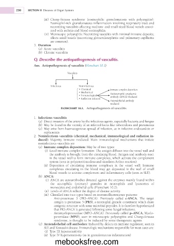
Q. Describe the aetiopathogenesis of vasculitis.
Ans. Aetiopathogenesis of vasculitis (Flowchart 10.1)
Vasculitis
Infectious Noninfectious
• Chemical Immune complex deposition
• Mechanical Antineutrophil cytoplasmic
• Immunological antibody (ANCA) mediated
• Radiation induced
Antiendothelial antibody
mediated
FLOWCHART 10.1. Aetiopathogenesis of vasculitis.
1. Infectious vasculitis
(a) Direct invasion of the artery by the infectious agents, especially bacteria and fungus
(b) May be found in the vicinity of an infected focus like tuberculosis and pneumonia
(c) May arise from haematogenous spread of infection, as in infective endocarditis or
septicaemia
2. Noninfectious vasculitis (chemical, mechanical, immunological and radiation in-
duced): Majority, immune mediated. Main immunological mechanisms that initiate
noninfectious vasculitis are
(a) Immune complex deposition: May be of two types:
(i) Local immune complex formation: The antigen diffuses into the vessel wall and
the antibody is brought from the circulating blood. Antigen and antibody react
in the vessel wall to form immune complexes, which activate the complement
system (seen in polyarteritisnodosa and simulates Arthus reaction).
(ii) Deposition of circulating immune complexes in the vessel wall: Immune
complexes circulating in the blood may get deposited in the wall of small
blood vessels to activate complement and inflammatory cells (seen in SLE).
(b) ANCA:
(i) ANCA are autoantibodies directed against the enzymes mainly found within
the azurophilic (primary) granules in neutrophils and lysosomes of
monocytes and endothelial cells (Flowchart 10.2).
(ii) Levels of ANCA reflect the degree of disease activity.
(iii) Classified into two types based on immunofluorescence patterns:
- Anti-proteinase 3 (PR3–ANCA): Previously called c-ANCA. The target
antigen is proteinase 3 (PR3), a neutrophil granule constituent which share
antigenic structure with some microbial peptides. It is therefore hypothesized
that PR3-ANCA is generated following some fungal infections.
- Antimyeloperoxidase (MPO–ANCA): Previously called p–ANCA, Myelo-
peroxidase (MPO); seen in microscopic polyangiitis and Churg–Strauss
syndrome, is thought to be induced by some therapeutic agents.
(c) Antiendothelial cell antibodies: Induced by defects in immune regulation; seen in
SLE and Kawasaki disease. Immunologic mechanisms responsible for most cases are:
(i) Type III hypersensitivity
(ii) Type IV hypersensitivity (as in granulomatous inflammation)
mebooksfree.com