
Page 357 - Concise Pathology for Exam Preparation ( PDFDrive )
P. 357
342 SECTION II Diseases of Organ Systems
Mature-
appearing
lymphocytes
Smudge cells
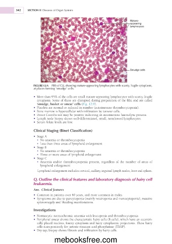
FIGURE 12.9. PBS of CLL showing mature-appearing lymphocytes with scanty, fragile cytoplasm,
at places forming ‘smudge’ cells.
• More than 95% of the cells are small mature-appearing lymphocytes with scanty, fragile
cytoplasm. Some of these are disrupted during preparation of the film and are called
‘smudge, basket or smear’ cells (Fig. 12.9).
• Platelets are normal or reduced in number (autoimmune thrombocytopenia).
• Bone marrow is hypercellular with infiltration by tumour cells.
• Direct Coombs test may be positive indicating an autoimmune haemolytic process.
• Lymph node biopsy shows well-differentiated, small, noncleaved lymphocytes.
• Serum folate levels are low.
Clinical Staging (Binet Classification)
• Stage A
• No anaemia or thrombocytopenia
• Less than three areas of lymphoid enlargement
• Stage B
• No anaemia or thrombocytopenia
• Three or more areas of lymphoid enlargement
• Stage C
• Anaemia and/or thrombocytopenia present, regardless of the number of areas of
lymphoid enlargement
Lymphoid enlargement includes cervical, axillary, inguinal lymph nodes, liver and spleen.
Q. Outline the clinical features and laboratory diagnosis of hairy cell
leukaemia.
Ans. Clinical features
• Common in patients over 40 years, and more common in males.
• Symptoms are due to pancytopenia (mainly neutropenia and monocytopenia), massive
splenomegaly and bleeding manifestations.
Investigations
• Normocytic normochromic anaemia with leucopenia and thrombocytopenia.
• Peripheral smear shows the characteristic hairy cells (B cells), which have an eccentri-
cally placed nucleus, foamy cytoplasm and hairy cytoplasmic projections. These hairy
cells stain positively for tartrate-resistant acid phosphatase (TRAP).
• Dry tap; biopsy shows fibrosis and infiltration by hairy cells.
mebooksfree.com