
Page 359 - Concise Pathology for Exam Preparation ( PDFDrive )
P. 359
344 SECTION II Diseases of Organ Systems
Plasma cells
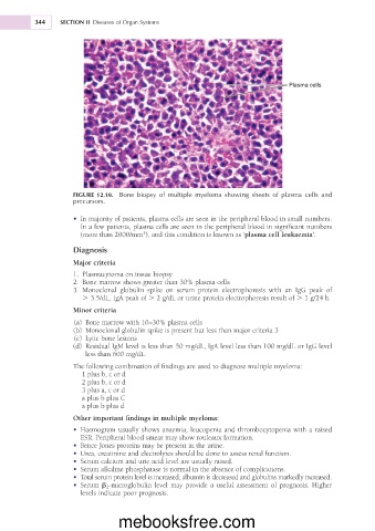
FIGURE 12.10. Bone biopsy of multiple myeloma showing sheets of plasma cells and
precursors.
• In majority of patients, plasma cells are seen in the peripheral blood in small numbers.
In a few patients, plasma cells are seen in the peripheral blood in significant numbers
(more than 2000/mm ), and this condition is known as ‘plasma cell leukaemia’.
3
Diagnosis
Major criteria
1. Plasmacytoma on tissue biopsy
2. Bone marrow shows greater than 30% plasma cells
3. Monoclonal globulin spike on serum protein electrophoresis with an IgG peak of
. 3.5/dL, IgA peak of . 2 g/dL or urine protein electrophoresis result of . 1 g/24 h
Minor criteria
(a) Bone marrow with 10–30% plasma cells
(b) Monoclonal globulin spike is present but less than major criteria 3
(c) Lytic bone lesions
(d) Residual IgM level is less than 50 mg/dL, IgA level less than 100 mg/dL or IgG level
less than 600 mg/dL.
The following combination of findings are used to diagnose multiple myeloma:
1 plus b, c or d
2 plus b, c or d
3 plus a, c or d
a plus b plus C
a plus b plus d
Other important findings in multiple myeloma:
• Haemogram usually shows anaemia, leucopenia and thrombocytopenia with a raised
ESR. Peripheral blood smear may show rouleaux formation.
• Bence Jones proteins may be present in the urine.
• Urea, creatinine and electrolytes should be done to assess renal function.
• Serum calcium and uric acid level are usually raised.
• Serum alkaline phosphatase is normal in the absence of complications.
• Total serum protein level is increased, albumin is decreased and globulins markedly increased.
• Serum b 2 -microglobulin level may provide a useful assessment of prognosis. Higher
levels indicate poor prognosis.
mebooksfree.com