
Page 617 - Concise Pathology for Exam Preparation ( PDFDrive )
P. 617
602 SECTION II Diseases of Organ Systems
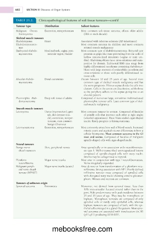
TABLE 21.2. Clinicopathological features of soft tissue tumours—cont’d
Tumour type Distribution Salient features
Malignant fibrous Extremities, retroperitoneum Most common soft tissue sarcoma; affects older adults
histiocytoma (fifth to sixth decade).
Skeletal muscle tumours
Rhabdomyoma Heart Associated with tuberous sclerosis (AD inheritance).
Rhabdomyosarco- Most common sarcoma in children and most common
mas striated muscle malignancy.
Embryonal rhabdo- Head and neck, vagina, para- Most common type of rhabdomyosarcoma. Botryoid type
myosarcoma testicular region, bladder presents as grape-like mass protruding from the walls of
hollow mucosa-lined structures (vagina or male ure-
thra). Rhabdomyoblasts have cross-striations and stain-
positive for desmin. Embryonal RMS may range from
highly differentiated neoplasms containing rhabdomyo-
blasts with large amounts of eosinophilic cytoplasm and
cross-striations to those with poorly differentiated tu-
mour cells.
Alveolar rhabdo- Distal extremities Occurs between 10 and 25 years of age. Second most
myosarcoma common type of skeletal muscle malignancy and has
the worst prognosis. Fibrous septae divide the cells into
clusters. Cells in the centre are discohesive, while those
at the periphery adhere to the septae giving rise to an
alveolar pattern.
Pleomorphic rhab- Deep soft tissue of adults Composed of numerous large, sometimes multinucleated
domyosarcoma pleomorphic tumour cells. Least common type of skel-
etal muscle malignancy.
Smooth muscle tumours
Leiomyoma Uterus (myometrium), geni- Most common tumour in women. Composed of fascicles
tals, skin (erector mus- of spindle cells that intersect each other at right angles
cle), extremities, retroper- (whorled appearance). Have blunt-ended cigar-shaped
itoneum, most common nuclei. Rarely progress to leiomyosarcoma.
benign GI tumour
Leiomyosarcoma Extremities, retroperitoneum Most commonly arises from wall of blood vessels. Increased
mitotic count and atypical mitoses differentiate it from a
cellular leiomyoma. Most common sarcoma in the GI
tract and uterus. Composed of fascicles of malignant
spindle-shaped cells with cigar-shaped nuclei.
Neural tumours
Benign nerve Skin, peripheral nerves Arise sporadically or in association with neurofibromato-
sheath tumours sis type I. Well-circumscribed unencapsulated lesions
composed of spindle-shaped cells with wavy nuclei.
Stroma may be collagenized to myxoid.
Plexiform Major nerve trunks Most arise in conjunction with type I neurofibromatosis.
neurofibroma Nerve irregularly expanded.
Malignant periph- Major nerve trunks (sciatic) Arise de novo or from transformation of a plexiform neu-
eral nerve sheath rofibroma. Strong association with NF I. Poorly defined
tumour (MPNST) infiltrative tumour mass composed of spindled cells
with elongated wavy nuclei showing extreme pleomor-
phism. Mitoses and necrosis are common.
Tumours of unknown origin
Synovial sarcoma Extremities Misnomer; not derived from synovial tissue. Less than
10% intra-articular. Located around rather than in the
joint. Male predominance with peak incidence between
25 and 35 years of age. They may be monophasic or
biphasic. Monophasic tumours are composed of only
spindled cells or rarely only epithelial cells, whereas
biphasic tumours are composed of both, with the epi-
thelial cells arranged in a gland-like pattern. Most syno-
vial sarcomas are associated with translocation t(x;18)
(p11;q11) producing SS18-SSX1.
mebooksfree.com