
Page 616 - Concise Pathology for Exam Preparation ( PDFDrive )
P. 616
21 Musculoskeletal System 601
Q. Write briefly on myasthenia gravis.
Ans. Autoimmune disease characterized by reduction in acetylcholine receptors due to
the presence of an autoantibody against them.
• Acetylcholine receptor antibody accelerates degradation of the receptor aided by
complement activation and blocks receptor function.
• Myasthenia gravis may be associated with thymic hyperplasia as well as thymomas.
• Ptosis and diplopia are the most common initial presentations.
• Histopathology is not diagnostic; Type II fibre atrophy may be observed in late stage.
• Treatment includes anticholinesterase drugs, thymectomy, immunosuppression and
plasmapheresis.
Q. Write briefly on Lambert–Eaton myasthenic syndrome.
Ans. Develops as a paraneoplastic process, commonly with small cell carcinoma of lung.
• Patients develop proximal muscle weakness with autonomic dysfunction.
• No improvement found with anticholinesterase agents.
• Content of anticholinesterase is normal in neuromuscular junction synaptic vesicles, but
fewer vesicles are released.
Soft Tissue
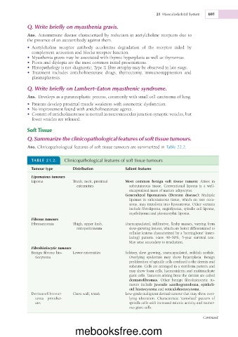
Q. Summarize the clinicopathological features of soft tissue tumours.
Ans. Clinicopathological features of soft tissue tumours are summarized in Table 22.2.
TABLE 21.2. Clinicopathological features of soft tissue tumours
Tumour type Distribution Salient features
Lipomatous tumours
Lipoma Trunk, neck, proximal Most common benign soft tissue tumour. Arises in
extremities subcutaneous tissue. Conventional lipoma is a well-
encapsulated mass of mature adipocytes.
Generalized lipomatosis (Dercum disease): Multiple
lipomas in subcutaneous tissue, which on rare occa-
sions, may transform into liposarcoma. Other variants
include fibrolipoma, angiolipoma, spindle cell lipoma,
myelolipoma and pleomorphic lipoma.
Fibrous tumours
Fibrosarcomas Thigh, upper limb, Unencapsulated, infiltrative, fleshy masses, varying from
retroperitoneum slow-growing lesions, which are better differentiated to
cellular lesions characterized by a ‘herringbone’ (inter-
lacing) pattern. Have 40–50%, 5-year survival rate.
May arise secondary to irradiation.
Fibrohistiocytic tumours
Benign fibrous his- Lower extremities Solitary, slow growing, unencapsulated, reddish nodule.
tiocytoma Overlying epidermis may show hyperplasia. Benign
proliferation of spindle cells confined to the dermis and
subcutis. Cells are arranged in a storiform pattern and
may show foam cells, haemosiderin and multinucleate
giant cells. Tumours arising from the dermis are called
dermatofibromas. Other benign fibrohistiocytic tu-
mours include juvenile xanthogranuloma, epitheli-
oid histiocytoma and reticulohistiocytoma.
Dermatofibrosar- Chest wall, trunk Low-grade malignant dermal tumour that may show over-
coma protuber- lying ulceration. Characteristic ‘cartwheel’ pattern of
ans spindle cells with increased mitotic activity and numer-
ous giant cells.
Continued
mebooksfree.com