
Page 1235 - Hematology_ Basic Principles and Practice ( PDFDrive )
P. 1235
Chapter 68 The Polycythemias 1081
Asymptomatic evaluation to differentiate this disorder from other causes of spurious
Splenomegaly or absolute erythrocytosis. The diagnosis of PV has been simplified
Isolated erythrocytosis by the identification of PV-associated mutations (JAK2V617F and
Isolated thrombocytosis JAK2 exon 12 mutations), permitting for the first time molecular
epidemiologic studies. The prevalence of the JAK2V617F mutation
in a normal population in Denmark was recently determined to be
0.2%, with 63% of these individuals not having a previously detected
Erythrocytotic phase hematological malignancy. The presence of the mutation was associ-
Erythrocytosis ated with increasing age, male sex, and a lower cumulative survival.
Thrombocytosis Recently available 2001–2012 data from the Surveillance, Epidemiol-
Leukocytosis ogy and End Results (SEER) Program from the US National Cancer
Splenomegaly Institute provided new insight into the IR of the MPNs. This study
Thrombosis utilized the WHO Diagnostic Criteria and for the last 5 years of the
Hemorrhage study JAK2V617F testing was included. The age adjusted IR of PV
Pruritus and essential thrombocythemia (ET) were 10.9 and 9.4 per 1 million
per year, respectively. The IR for PMF was 3.1. In order to grasp the
magnitude of the IR of these various MPNs, the IR of CML was 3.3,
−
demonstrating that the IR of these Ph MPNs were sevenfold higher
Post-PV MF, myelofibrosis than CML. Both PV and PMF were more frequent in males while
Anemia the IR for ET was greater in females. Several groups have reported
Leukoerythroblastosis
Thrombocytopenia or that 50% of patients with a splanchnic vein thrombosis without an
thrombocytosis overt MPN are JAK2V617F positive and that more than 50% of these
Enlarging splenomegaly individuals subsequently develop an MPN. However, the incidence
Systematic symptoms of the mutation with unprovoked thromboembolism in the more
(fever, weight loss) usual sites (deep venous thrombosis involving the leg veins or pul-
monary embolism) ranges between 0.2 and 1.0%. Such data have led
to the conclusion that systematic screening for JAK2V617 in such a
patient population is not warranted.
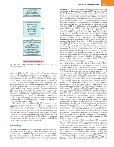
Acute myeloid leukemia The prevalence of PV has been reported by several investigators
to be higher among American Jews and lower among African–
Fig. 68.3 EVOLUTION OF POLYCYTHEMIA VERA. MF, Myelofibro- Americans and Hispanics. The reported lower incidence in African–
sis; PV, polycythemia vera.
Americans might reflect a referral bias characteristic of centers with
research interest in MPN because the authors, when practicing in
several large urban areas, have observed a considerable number of
such as myelofibrosis (MF), termed post-PV MF, and acute leukemia African–Americans with MPNs, including PV. The incidence of the
(Fig. 68.3). Frequently, patients present asymptomatically to a physi- disorder is greater among Ashkenazi Jews, who originate from eastern
cian only to find that they have splenomegaly, isolated erythrocytosis, and central Europe, than among Arabs and Sephardic Jews. Interest-
or thrombocytosis. Left untreated, these patients will become symp- ingly, extremely low occurrence rates have been reported from Japan.
tomatic owing to the excessive production of RBCs, platelets, or These findings suggest that important genetic factors might be
both, leading to arterial or venous thromboses, aquagenic pruritus, involved in the biogenesis of this disorder. The importance of genetic
and symptoms caused by increasing splenomegaly. After a number of factors in the origin of this disease is further emphasized by reports
years, the erythrocytotic phase of the disease frequently becomes of multiple cases of JAK2V617F or JAK2 exon 12 mutation-positive
inactive, and the patient may no longer have the sequelae of excessive and -negative MPN, including PV, within multiple generations of a
RBC production. Subsequently, these patients can develop post-PV number of families. These forms of familial PV must be distinguished
MF, which is frequently indistinguishable from another MPN, from PFCP, CP, and polycythemia associated with mutations in the
primary MF (PMF). Finally, a significant proportion of these patients HIF pathway. The reports of families in which multiple members
will go on to develop acute myeloid leukemia (AML). Only a limited have PV first raised the possibility that a genetic predisposition to
number of patients undergo this orderly transition; many patients acquire such mutations exists in these families that is inherited in an
transition from the polycythemic phase directly into an acute leuke- autosomal dominant pattern with decreased penetrance. Clinical
mia or a myelodysplastic disorder. 9 analyses of affected family members confirmed that they have clonal
The transition from one phase of this MPN to another is not hematopoiesis and that their clinical manifestations are identical to
necessarily unidirectional. About 10–15% of patients with patients with sporadic PV. In a large population study from Sweden,
JAK2V617F-positive essential thrombocythemia eventually develop the ratives of MPN patients were shown to have a significantly
−
erythrocytosis and are reclassified as having PV. A number of cases of increased risk of developing a Ph MPN and possibly chronic myeloid
10
presumed PMF either spontaneously or after chemotherapy have leukemia. It was estimated that first-degree relatives of MPN
been described where the patient develops erythrocytosis and a syn- patients have a five- to sevenfold greater risk of developing an MPN,
drome that is virtually indistinguishable from de novo PV. The again supporting the hypothesis that common strong susceptibility
constantly changing clinical picture of this malignant hematologic genes predispose one to develop PV, essential thrombocytosis (ET),
disorder requires careful observation and treatment to deal with the PMF, and possibly CML.
numerous problems that can be encountered. One notable exception to the low prevalence of PV in Japan has
been the higher incidence observed among populations exposed to
atomic bomb explosions. The possibility that radiation exposure is
Epidemiology an etiologic factor in the generation of PV was also raised by the
observation in the United States of four cases of PV 10–20 years after
PV is the most common primary polycythemia. Incidence rates (IRs) the Smokey nuclear weapons test in which 3000 military observers
have been reported to be around 2.8 per 100,000 persons per year. were exposed. An epidemiologic investigation that focused on occu-
It is important to emphasize that no or very few population-based pational exposure among petroleum refinery and chemical plant
estimates of the prevalence of this disorder are presently available. workers has revealed an increased incidence of PV relative to the
Actual determination of its prevalence has been a difficult process general population. In this study, the increased incidence of PV was
because of the need in the past to pursue an extensive diagnostic linked to similar increases in the frequency of multiple myeloma and