
Page 162 - Hematology_ Basic Principles and Practice ( PDFDrive )
P. 162
120 Part II Cellular Basis of Hematology
9
providing signals required for maintenance, quiescence and retention HSPC number further supporting the notion of a perivascular niche.
of HSPCs in the BM. However, the location of HSPC niche within Although the debate about location of the HSPC niche, and conse-
6
the marrow has been a subject of controversy. The endosteal surface quently, cell types that serve as niche participants, continues it is
has long been considered the zone in which HSPCs are preferentially important to bear in mind that HSPCs themselves are molecularly
located. In the setting of irradiation conditioning, this has been and functionally heterogeneous, and that several distinct niches may
directly demonstrated by intravital imaging studies, which allow coexist to support this heterogeneity, particularly under different
dynamic assessment of the interaction between transplanted HSPC conditions such as the stress of transplantation.
and the niche. Currently, in vivo imaging is limited to calvarial BM,
an area in the mouse skull where the bone is very thin, thus permit-
ting penetration of the laser beam into the BM cavity. Using this Cellular Components of the HSPC Niche
technique and simultaneous multicolor fluorescent labeling of osteo-
lineage cells (OLCs), HSPCs, and the vasculature, studies showed Over recent years, animal studies revealed marked complexity in
that in irradiated recipients, transplanted HSPCs home closest to the cellular and molecular organization of the HSPC BM niche. Major
endosteal surface and individual OLCs as compared with more dif- cellular components of the HSPC niche and the factors that they
ferentiated progenitors, and that they are “anchored” to their niches produce (summarized in Table 11.1) are discussed later.
7
at least through 72 hours. Preferential localization of primitive
hematopoietic cells to the endosteal surface under the homeostatic
conditions has been also demonstrated, although this analysis was Osteolineage Cells
performed using immunostaining of histologic BM sections of either
femoral bones or the sternum. 8 OLCs are a heterogeneous population of mesenchymal cells that line
However, other studies performed under steady state (not trans- the endosteal surfaces of flat and trabeculated bones at the interface
plant) conditions indicate that most HSPC are located in the central between the bone and the BM and become embedded within
marrow in a perivascular position, thus arguing in favor of more primi- the bone matrix upon terminal differentiation. OLCs are thought
tive mesenchymal cells and endothelial cells governing the niche. to originate from mesenchymal stem cells (MSCs) and gradually
Deletion of a key niche factor, such as stem cell factor (kit-ligand), progress from the early immature progenitors that express OLC-
from either endothelial or perivascular cells leads to decrease in the specific transcription factors Runx2 and osterix to mature osteoblasts
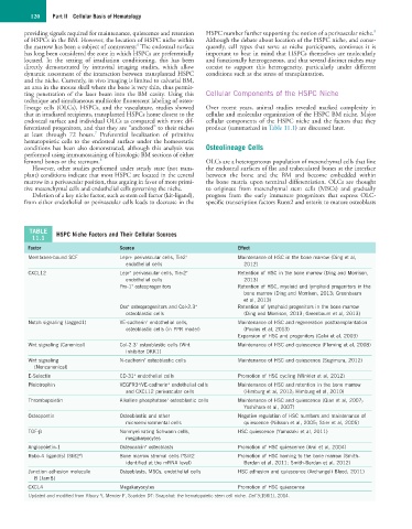
TABLE HSPC Niche Factors and Their Cellular Sources
11.1
Factor Source Effect
+
Membrane-bound SCF Lepr+ perivascular cells, Tie2 Maintenance of HSC in the bone marrow (Ding et al,
endothelial cells 2012)
+
CXCL12 Lepr perivascular cells, Tie-2 Retention of HSC in the bone marrow (Ding and Morrison,
+
endothelial cells 2013)
+
Prx-1 osteoprogenitors Retention of HSC, myeloid and lymphoid progenitors in the
bone marrow (Ding and Morrison, 2013; Greenbaum
et al, 2013)
+
+
Osx osteoprogenitors and Col-2.3 Retention of lymphoid progenitors in the bone marrow
osteoblastic cells (Ding and Morrison, 2013; Greenbaum et al, 2013)
+
Notch signaling (Jagged1) VE-cadherin endothelial cells, Maintenance of HSC and regeneration posttransplantation
osteoblastic cells (in PPR model) (Poulos et al, 2013)
Expansion of HSC and progenitors (Calvi et al, 2003)
+
Wnt signaling (Canonical) Col-2.3 osteoblastic cells (Wnt Maintenance of HSC and quiescence (Fleming et al, 2008)
inhibitor DKK1)
+
Wnt signaling N-cadherin osteoblastic cells Maintenance of HSC and quiescence (Sugimura, 2012)
(Noncanonical)
+
E-Selectin CD-31 endothelial cells Promotion of HSC cycling (Winkler et al, 2012)
+
+
Pleiotrophin VEGFR3 /VE-cadherin endothelial cells Maintenance of HSC and retention in the bone marrow
and CXCL12 perivascular cells (Himburg et al, 2012; Himburg et al, 2010)
+
Thrombopoietin Alkaline phosphatase osteoblastic cells Maintenance of HSC and quiescence (Qian et al, 2007;
Yoshihara et al, 2007)
Osteopontin Osteoblastic and other Negative regulation of HSC numbers and maintenance of
microenvironmental cells quiescence (Nilsson et al, 2005; Stier et al, 2005)
TGF-β Nonmyelinating Schwann cells, HSC quiescence (Yamazaki et al, 2011)
megakaryocytes
Angiopoietin-1 Osteocalcin osteoblasts Promotion of HSC quiescence (Arai et al, 2004)
+
a
a
Robo-4 ligand(s) (Slit2 ) Bone marrow stromal cells ( Slit2 Promotion of HSC homing to the bone marrow (Smith-
identified at the mRNA level) Berdan et al, 2011; Smith-Berdan et al, 2012)
Junction adhesion molecule Osteoblasts, MSCs, endothelial cells HSC adhesion and quiescence (Archangeli Blood, 2011)
B (JamB)
CXCL4 Megakaryocytes Promotion of HSC quiescence
Updated and modified from Kfoury Y, Mercier F, Scadden DT: Snapshot: the hematopoietic stem cell niche. Cell 3;158(1), 2014.