
Page 2078 - Hematology_ Basic Principles and Practice ( PDFDrive )
P. 2078
Chapter 123 The Blood Vessel Wall 1847
is dependent on multiple attractive and repulsive cues, many of which lumen diameter and wall thickness, requires both cell death and
are common to both the nervous and vascular systems. 112,113 Tip cells proliferation (as well as remodeling of the ECM). In addition to
express a distinctive profile of genes with substantially higher expres- survival signals transmitted by integrins, shear stress is important for
sion compared with stalk cells of molecular markers including endothelial survival and vessel healing after injury. 117–119 Oxygen
platelet-derived growth factor (PDGF)-B, VEGFR-2, uncoordinated tension is important in vascular maintenance. Hypoxia increases
(Unc)5b, Delta-like (Dll)4, and VEGFR-3. Whereas VEGF165 acts levels of VEGF, which provides signals for vessel maintenance and
120
as an attractive cue to the tip cell of the endothelial sprout, Netrin-1 neovascularization. Hyperoxia, on the other hand, inhibits VEGF
121
signals to Unc5b on the vasculature act as a repulsive cue. Netrin-4 expression, which leads to regression and death of retinal vessels.
can also bind Neogenin, which in turn recruits and activates Unc5b In some models, regression of vessels occurs by apoptosis of vascular
to mediate repulsion. Other guidance pathways implicated in vascular cells. 122,123 Endothelial cells express several antiapoptotic molecules to
patterning and angiogenesis are ephrinB2–EphB4, plexinD1– maintain viability when quiescent and when stressed. 124,125 Most
semaphorin, and Slit–Robo interactions, as well as the neuropilins. likely, an intricate balance between cell death and proliferation is
Patterning and specification of small arteries along peripheral nerves maintained by activators and inhibitors of both processes.
in the skin of the embryonic limb involves nerve-derived VEGF; in
other situations, neuronal patterning is dependent on the vascula-
ture. 114,115 Thus the congruent patterning of the neural and vascular Role of Ligand–Receptor Interactions
systems likely is caused by use of common signals and may require
cross-talk between the two systems. Numerous factors regulate vascular development and differentiation
in a positive or negative fashion. Some of the key molecules and their
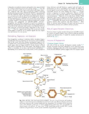
receptors are discussed here. A model for vascular development is
Remodeling, Regression, and Apoptosis shown in Fig. 123.1.
Even though the vasculature is laid down before circulation begins,
hemodynamic forces are important for maintenance and remodeling. Inducers of Angiogenesis
Most of the vessels laid down during vasculogenesis regress or are
remodeled. After neovascularization (e.g., during wound healing), the Fibroblast Growth Factors
vessels regress when no longer needed. A chronic decrease in blood The role of FGFs in vascular development remains murky. 126–128
flow results in narrowing of the vessel lumen. This change in vessel Because of possible functional redundancy in the numerous family
116
caliber is dependent on an intact and functional endothelium. members, assigning specific roles to the various members of the FGF
Remodeling, which involves loss of some vessels as well as changes in family has been difficult. Evidence suggests that FGF receptors signal
Vasculogenesis Angiogenesis
Blood islands
Ectoderm PEC
Angioblast
Mesoderm
FGF2 VEGFR-2
Endoderm VEGF VEGF-R2/R1 Hematopoietic Basement
precursor membrane
VEGF-R2/R1 Tie2
VEGF-R2/R1
Ang2
Capillary
plexus VEGF
PDGF-Rb
PDGF-BB
TF?
Mesenchymal
Recruitment of PEC cell
Ang1
Tie2
TGFβ
Quiescence Regression/apoptosis
PEC
PEC
Inhibition of EC proliferation TF?
PEC differentiation
ECM depositiion Apoptotic EC
Tube formation
ECM Tie2 Ang2
Fig. 123.1 MODEL FOR VASCULAR DEVELOPMENT. The role of secreted proteins and membrane
receptors in vascular development is highlighted, but other factors such as cell adhesion molecules and extracel-
lular matrix components also contribute significantly. Ang, Angiopoietin; EC, extracellular; ECM, extracellular
matrix; FGF2, fibroblast growth factor 2; PDGF, platelet-derived growth factor; PEC, periendothelial cell
(smooth muscle cell, pericyte); TF, tissue factor; TGFβ, transforming growth factor-β; VEGF, vascular
endothelial cell growth factor; VEGFR, vascular endothelial cell growth factor receptor.