
Page 2198 - Hematology_ Basic Principles and Practice ( PDFDrive )
P. 2198
Chapter 131 Diseases of Platelet Number 1945
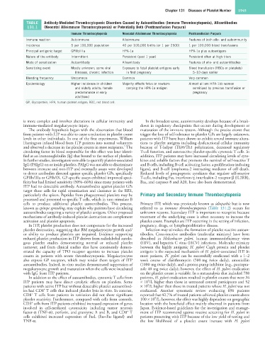
TABLE Antibody-Mediated Thrombocytopenic Disorders Caused by Autoantibodies (Immune Thrombocytopenia), Alloantibodies
131.1 (Neonatal Alloimmune Thrombocytopenia) or Potentially Both (Posttransfusion Purpura)
Immune Thrombocytopenia Neonatal Alloimmune Thrombocytopenia Posttransfusion Purpura
Immune reaction Autoimmune Alloimmune Features of both allo- and autoimmunity
Incidence 5 per 100,000 population 40 per 100,000 births (or 1 per 2500) 1 per 100,000 blood transfusions
Principal antigenic target GPIIb/IIIa HPA-1a HPA-1a plus autoantigens
Nature of the antibody Intermittent Persistent (past 1 year) Persistent often at high titers
Mode of sensitization Autoantibody Alloantibody Features of allo- and autoantibodies
Sensitizing event Mostly unknown; some viral Exposure to fetal platelet antigens early Blood transfusion (RBCs or platelets)
illnesses, chronic infection in first pregnancy 5–10 days earlier
Bleeding frequency Uncommon Common Very common
Epidemiology Higher incidence in children Majority affects fetus or newborn Almost all are HPA-1bb women
and elderly adults; female carrying the HPA-1a antigen sensitized by previous transfusion or
predominance in early pregnancy
adulthood
GP, Glycoprotein; HPA, human platelet antigen; RBC, red blood cell.
is more complex and involves alterations in cellular immunity and In the broadest sense, autoimmunity develops because of a break-
immune-mediated megakaryocyte injury. down in regulatory checkpoints that occurs during development or
The antibody hypothesis began with the observation that blood maturation of the immune system. Although the precise events that
from patients with ITP was able to cause a reduction in platelet count trigger the loss of self-tolerance to platelet GPs are largely unknown,
levels in other individuals. In one of the first experiments, William patients with ITP have been shown to exhibit several immune altera-
Harrington infused blood from ITP patients into normal volunteers tions to platelet antigens including dysfunctional cellular immunity
5
and observed a decrease in the platelet counts in most recipients. The because of T-helper (Th)0/Th1 polarization, decreased regulatory
circulating factor in blood responsible for this effect was later identi- T-cell function, and autoreactive platelet-specific cytotoxic T cells. In
fied as an immunoglobulin (Ig) that bound to the surface of platelets. addition, ITP patients may have increased circulating levels of cyto-
In further studies, investigators were able to quantify platelet-associated kines and soluble factors that promote the survival of self-reactive T
IgG (PAIgG) on or inside platelets. PAIgG was not able to discriminate and B cells, including B-cell activating factor, a proliferation-inducing
between immune and non-ITP and eventually assays were developed ligand, and B-cell lymphoma-2 interacting mediator of cell death.
to detect antibodies directed against specific platelet GPs, specifically Reduced levels of proapoptotic cytokines that regulate self-reactive
GPIIb/IIIa or GPIb/IX. GP-specific assays exhibited improved speci- T-cells, including Fas, interferon-γ, interleukin-2 receptor β (IL2RB),
ficity but had limited sensitivity (50%–66%) since many patients with Bax, and caspases 8 and A20, have also been demonstrated.
ITP had no detectable antibody. Autoantibodies against platelet GPs
target those cells for rapid opsonization and clearance in the RES,
particularly the spleen. Peptides from phagocytosed platelets may be Primary and Secondary Immune Thrombocytopenia
processed and presented to specific T cells, which in turn stimulate B
cells to produce additional platelet autoantibodies. This process, Primary ITP, which was previously known as idiopathic but is now
known as epitope spreading, may explain why patients have circulating referred to as immune thrombocytopenia (Table 131.2) occurs for
autoantibodies targeting a variety of platelet antigens. Other proposed unknown reasons. Secondary ITP is important to recognize because
mechanisms of antibody-induced platelet destruction are complement treatment of the underlying cause is often necessary to increase the
activation and platelet apoptosis. platelet count. Examples are ITP occurring in the setting of infection,
In ITP, platelet production does not compensate for the increased pregnancy, drugs, or lymphoproliferative disease.
platelet destruction, suggesting that BM megakaryocyte growth and/ Infection may stimulate the formation of platelet reactive autoan-
or ability to produce platelets are impaired. Evidence supporting tibodies. Cross-reactive antibodies (molecular mimicry) have been
reduced platelet production in ITP derives from radiolabeled autolo- described in Helicobacter pylori, human immunodeficiency virus
gous platelet studies demonstrating normal or reduced platelet (HIV), and hepatitis C virus (HCV) infections. Molecular mimicry
turnover; and from clinical studies that have consistently demon- between the highly antigenic H. pylori CagA protein and platelet
strated the capacity of TPO receptor agonists to increase platelet antigens is the suspected mechanism of H. pylori–associated ITP. In
counts in patients with severe thrombocytopenia. Megakaryocytes most patients, H. pylori can be successfully eradicated with a 1–2
also express GP receptors, which may render them targets of ITP week course of clarithromycin (500 mg twice daily), amoxicillin
autoantibodies. Indeed, in vitro studies demonstrated suppression of (1000 mg twice daily), and a proton pump inhibitor (e.g., pantopra-
megakaryocyte growth and maturation when the cells were incubated zole 40 mg twice daily); however, the effect of H. pylori eradication
with IgG from ITP patients. on the platelet count is variable. In a metaanalysis that included 788
In addition to the effect of autoantibodies, cytotoxic T cells from patients, H. pylori eradication resulted in platelet counts that were 34
9
ITP patients may have direct cytolytic effects on platelets. Some × 10 /L higher than those in untreated control participants and 52
9
patients with active ITP but without detectable platelet autoantibod- × 10 /L higher than those in treated patients whose H. pylori was not
+
ies had CD8 T cells that induced platelet lysis in vitro. In contrast, eradicated. Another systematic review evaluating 696 patients
+
CD8 T cells from patients in remission did not show significant reported that 42.7% of treated patients achieved platelet counts above
9
platelet reactivity. Furthermore, compared with cells from controls, 100 × 10 /L; however, the effect was highly dependent on geographic
+
CD3 cells from ITP patients exhibited increased expression of genes location with the beneficial effect mainly observed in patients from
involved in cell-mediated cytotoxicity including tumor necrosis Japan. Evidence-based guidelines for the investigation and manage-
+
factor-α (TNF-α), perforin, and granzyme A and B, and CD8 T ment of ITP recommend against routine screening for H. pylori in
cells exhibited increased expression of FasL (Fas–Fas ligand) and patients presenting with ITP because of the low yield of testing and
TNF-α. the low likelihood of a platelet count increase with H. pylori