
Page 2365 - Hematology_ Basic Principles and Practice ( PDFDrive )
P. 2365
Chapter 142 Venous Thromboembolism 2107
ventilated but not perfused) has a positive predictive value for PE of A low clinical pretest probability combined with a normal D-dimer
more than 90%, obviating the need for additional testing. excludes PE. Patients with intermediate or high pretest probability
The relative ease and accessibility of CT scanning has reduced the should undergo further testing. A negative helical CTPA or a normal
use of V/Q scanning for the diagnosis of PE. However, V/Q scanning perfusion lung scan result rules out clinically significant PE, and
is frequently preferred in two patient populations: young patients and anticoagulant therapy can be withheld. If an intraluminal filling
those with impaired renal function. CT scanning exposes patients to defect is seen on helical CTPA or the perfusion scan demonstrates
a higher dose of radiation than V/Q scanning, raising concern regard- one or more segmental (or larger) defects and ventilation to these
ing the risk of subsequent cancer. Although this risk varies depending regions is normal, a diagnosis of PE is made. Although a V/Q mis-
on a number of factors, concern about increasing the risk of breast match supports a diagnosis of PE, a V/Q “match” does not exclude
cancer in young women has prompted use of V/Q scanning prefer- PE, and further objective testing is required in these patients. Simi-
entially in this population. In patients with impaired renal function, larly, the diagnosis of PE cannot be excluded in patients with small
contrast dye administration for the CT scan can induce contrast perfusion defects (one or more subsegmental defects) or those with
nephropathy, increasing mortality up to 30% after such a procedure. indeterminate lung scan findings (in which the perfusion defects
V/Q lung scanning should ideally be reserved for patients with correspond with abnormalities on the chest radiograph). In these
normal chest radiographs because preexisting lung disease may result patients, venous ultrasonography should be performed. If DVT is
in indeterminate scans. documented, a diagnosis of PE can be assumed, and anticoagulant
Patients with large perfusion defects (involving one or more seg- therapy should be started. However, if results on these tests are nega-
ments or more extensive defects) and a V/Q mismatch have a 90% tive, additional objective investigations (e.g., pulmonary angiography)
probability of PE. Patients with a normal perfusion scan have less are required in patients with a high clinical pretest probability. For
than a 2% probability of having PE, excluding the diagnosis. However, those with a lower pretest probability of PE, an alternative strategy
most patients who have V/Q scanning performed will have neither is to withhold anticoagulants and to perform serial noninvasive tests
of these findings; rather, they will have either matched defects or small to detect venous thrombosis.
perfusion defects (indeterminate scan). Patients with these findings
require further investigation with either pulmonary angiography or
objective tests for DVT of the lower extremities. A patient with DIAGNOSIS OF ACUTE RECURRENT VENOUS
suspected PE, an indeterminate V/Q scan result, and positive findings THROMBOEMBOLISM
on compression ultrasound examination of the lower extremities can
be assumed to have PE. A patient with suspected PE, an indetermi- The diagnosis of acute recurrent VTE is challenging because the
nate scan, and a negative result on leg compression ultrasound clinical manifestations of recurrence are nonspecific. In addition,
examination requires additional testing (e.g., serial compression there are no clinical prediction rules specifically validated for patients
ultrasound examination after 7 days) because the thrombus may have with suspected recurrence, and diagnostic tests for acute VTE have
completely embolized to the lungs. limitations in this setting. Following treatment of acute DVT, incom-
plete resolution of thrombosis may be evident as chronic venous
occlusion on compression ultrasound, making it difficult to identify
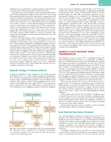
Diagnostic Strategy for Pulmonary Embolism new abnormalities. With respect to PE, the interpretation of repeat
CTPA or V/Q scanning may be difficult because of thrombus migra-
A diagnostic algorithm for the management of clinically suspected tion and variable rates of clot resolution. Therefore comparison with
PE is shown in Fig. 142.2. After a history and physical examination, prior imaging can facilitate diagnosis of acute recurrence. D-dimer
ECG, and chest radiography, the clinical probability of PE should be testing has limited utility in this setting because of the high frequency
assessed using a validated clinical prediction rule. The Wells clinical of positive D-dimer results in patients with suspected recurrence.
prediction rule assigns a pretest clinical probability based on the When assessing patients for recurrence, the adequacy of antico-
presence of the following: clinical signs of DVT, recent immobiliza- agulant therapy is an important consideration (e.g., nonadherence,
tion or surgery, heart rate above 100 beats/min, previous history of subtherapeutic international normalized ratio [INR] measurements,
VTE, hemoptysis, active malignancy, absence of alternative diagnosis. temporary interruption for procedure or surgery). Recurrent throm-
bosis despite adequate therapeutic anticoagulation can be seen in
patients with cancer or those with the antiphospholipid antibody
syndrome.
Clinically suspected PE Many patients with a history of VTE will have a heightened level
of concern about the risk of recurrent thrombosis, prompting them
to seek testing for recurrent thrombosis even with trivial symptoms.
Such patients require careful clinical and radiologic evaluation. If
Ventilation/perfusion there is no evidence of acute recurrence, they may require counseling
(V/Q) scan or helical and education about their condition.
CT scan Patients with suspected recurrence should be treated empirically
with anticoagulant therapy pending investigations.
Normal V/Q scan High-probability
V/Q scan or
Normal CT helical CT scan Acute Recurrent Deep Venous Thrombosis
Indeterminate The optimal diagnostic strategy for suspected recurrent DVT is
V/Q scan uncertain. The clinical history may be helpful in determining the
likelihood of recurrent thrombosis. Leg pain with ambulation and leg
swelling that is relieved with overnight rest is typical of the post-
Diagnosis excluded:
No further tests − Serial + Treat thrombotic syndrome. New clinically significant and persistent leg
indicated CUS swelling, particularly if the symptoms do not abate overnight, is
consistent with recurrent thrombosis and should prompt diagnostic
Fig. 142.2 DIAGNOSTIC ALGORITHM FOR THE MANAGEMENT evaluation.
OF PATIENTS WITH SUSPECTED PULMONARY EMBOLISM. To establish a diagnosis of recurrent DVT, one must demonstrate
CT, Computed tomography; CUS, compression ultrasound; PE, pulmonary a new thrombus in a previously unaffected venous segment. A normal
embolism V/Q, ventilation/perfusion. result on ultrasound study does not rule out recurrent calf vein