
Page 2488 - Hematology_ Basic Principles and Practice ( PDFDrive )
P. 2488
2220 Part XIII Consultative Hematology
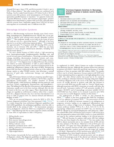
elevated fibrinogen, factor VIII, and fibrinopeptide A levels in up to Preliminary Diagnostic Guidelines for Macrophage
96
50% of these patients, but other studies have not confirmed such TABLE Activation Syndrome in Systemic Juvenile Idiopathic
findings. Another study found elevation of D-dimer levels in 96% of 152.2 Arthritis a
systemic-onset JIA patients; serial measurements of D-dimer levels
97
appeared to parallel response to treatment. In apparent distinction, Laboratory Criteria
decreased fibrinolytic activity and increased plasminogen activator 1. Decreased platelet count (≤262 × 10 /L)
9
inhibitor have been found in patients with active JIA, especially those 2. Elevated levels of aspartate aminotransferase (>59 IU/L)
with the systemic form. Antibodies against factor VIII and the lupus 3. Decreased white blood cell count (≤4.0 × 10 /L)
9
anticoagulant are occasionally seen in children with JIA. 4. Hypofibrinogenemia (≤250 mg/dL)
Clinical Criteria
Macrophage Activation Syndrome 1. Central nervous system dysfunction (irritability, disorientation,
lethargy, headache, seizures, coma)
2. Hemorrhages (purpura, easy bruising, mucosal bleeding)
MAS is a life-threatening multisystem disorder most closely resem- 3. Hepatomegaly (≥3 cm below the costal margin)
bling hemophagocytic lymphohistiocytosis (HLH) that occurs pri- Histopathologic Criterion
marily in patients with systemic-onset juvenile idiopathic arthritis
(sJIA). 98,99 This syndrome usually occurs early in the course of active Evidence of macrophage hemophagocytosis in the bone marrow aspirate
100
sJIA and can even be a presenting feature, although it has been Diagnostic Rule
101
reported to occur later and during a quiescent phase. It is estimated The diagnosis of macrophage activation syndrome requires the presence
that approximately 7% of patients with sJIA develop MAS, and the of any two or more laboratory criteria or of at least two clinical or
102
mortality rate is between 10% and 20%. MAS has also been laboratory criteria. A bone marrow aspirate for the demonstration of
reported in other systemic inflammatory disorders, including SLE hemophagocytosis may be required only in doubtful cases.
and Kawasaki disease. 100 a The suggested criteria are useful only in patients with active systemic-onset
The main clinical features of MAS include a high unremitting juvenile idiopathic arthritis. The laboratory thresholds are examples only and are
fever, hepatosplenomegaly, lymphadenopathy, bleeding, rash, and not specific for the diagnosis (see text).
From Ravelli A, Magni-Manzoni S, Pistorio A, et al: Preliminary diagnostic
central nervous system manifestations. Neurologic symptoms include guidelines for macrophage activation syndrome complicating systemic juvenile
lethargy, irritability, disorientation, headache, seizures, and coma. idiopathic arthritis. J Pediatr 146:598, 2005.
Children with MAS are acutely ill, and almost 50% require intensive
103
care unit care. The diagnosis may be delayed because the presenta-
tion mimics an acute exacerbation of sJIA or severe infection. Of
interest, some patients have shown a paradoxical improvement in the be emphasized. In MAS, clinical features are weaker discriminators
underlying inflammatory disease at the onset of MAS. Precipitating than laboratory features. Although the presence of fever was universal
factors that have been implicated include a flare-up of the underlying in patients with MAS, it had a low specificity rate because of the high
disease, aspirin or other NSAID toxicity, viral infections, a second incidence of fever in patients with sJIA without MAS. The pattern
injection of gold salts, methotrexate therapy, and sulfasalazine of fever may be of more importance because patients with MAS tend
therapy. 103 to have nonremitting fever as opposed to the high spiking fevers seen
Typical laboratory features include pancytopenia, hypofibrinogen- in sJIA. The other clinical criteria may occur late in the course of
emia (<250 mg/dL), elevated liver enzymes (>40 IU/mL), hypertri- MAS, resulting in abnormal laboratory findings being more helpful
glyceridemia (>160 mg/dL), and marked elevation of ferritin in making a diagnosis early in the course of the illness. Because blood
(>10,000 ng/mL). Other laboratory findings with less sensitivity and counts are usually elevated in patients with sJIA, the decrease in
specificity include coagulopathy, hyponatremia, and hypoalbumin- counts seen with MAS may actually result in a “normal” blood count,
emia. The hallmark of this disorder is the presence of hemophagocytic although in patients with HLH, cytopenias are usually well below the
histiocytes, usually seen in the bone marrow, although they can also normal levels. Laboratory markers of T-cell activation (soluble IL-2
be found in the liver and other organs. These characteristic macro- receptor [SCD25]) and of macrophage activation (soluble CD163
phages are regarded as confirmatory evidence rather than a require- [SCD163]) have been shown to be elevated in patients with MAS
ment for a diagnosis because they have not been documented in as and may serve as useful diagnostic markers in the future. 107
many as 20% of patients with the classical clinical and laboratory The pathogenesis of MAS is similar to that proposed for
findings of MAS. 103,104 HLH. 106,108 Indeed, it has been proposed that MAS be considered
102
There are no accepted diagnostic criteria for MAS in sJIA. Pre- one of the subtypes of acquired HLH. The presentation of MAS
104
liminary diagnostic guidelines have been proposed (Table 152.2). is a result of an ineffective immune response to an endogenous or
In a study comparing these preliminary diagnostic guidelines with exogenous stimulus leading to an exaggerated inflammatory state
HLH-2004 guidelines, the preliminary guidelines were best able to produced by a release of high levels of cytokines. These proinflam-
105
identify MAS in the setting of sJIA. An international consensus matory cytokines include TNF-α, IL-1, IL-6, IL-8, IL-12, IL-18,
survey of diagnostic criteria for MAS was completed by 232 physi- macrophage inflammatory protein-1α (MIP-1-α), and interferon-γ
105
cians. The following were the top nine diagnostic criteria identified (IFN-γ) released by stimulated lymphocytes and histiocytes. Defec-
by more 50% of respondents in order of frequency: tive natural killer (NK) cell function and cytotoxic T-cell activity have
been documented in patients with MAS as well as in those with HLH
1. Falling platelet count and may be the common pathway leading to the clinical presenta-
109
2. Hyperferritinemia tion. NK-cell function in patients with active sJIA but without
110
3. Bone marrow hemophagocytosis MAS has also been found to be abnormal. This may explain why
4. Increased live enzymes MAS is almost exclusively seen in the systemic form of JIA and not
5. Falling leukocyte count the other subtypes of the disease. Mutations in the perforin gene and
6. Persistent continuous fever >38°C the MUNC 13-4 gene have been described in some patients with
7. Falling erythrocyte sedimentation rate HLH. The cytotoxic function of NK cells is mediated by the release
8. Hypofibrinogenemia of perforin and other cytolytic granules into the target cell, leading
9. Hypertriglyceridemia to cell death. Abnormalities of both perforin gene and MUNC 13-4
polymorphisms have been found in patients with MAS. 109,111,112
These features may provide the best candidates for future refinement Treatment of MAS should be started promptly and not delayed
of diagnostic criteria. The diagnostic guidelines are similar to criteria for lack of hemophagocytosis if the clinical and laboratory features
106
for the diagnosis of HLH, although a number of differences should are consistent with the diagnosis. The initial treatment should be