
Page 363 - Hematology_ Basic Principles and Practice ( PDFDrive )
P. 363
Chapter 26 Biology of Erythropoiesis, Erythroid Differentiation, and Maturation 299
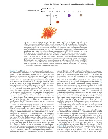
GMP CFU-GM
Stem cell CMP
MEP BFU-E CFU-E Proerythroblast Red cell
In vitro In vitro
14 days 7 days
Fig. 26.1 CELLULAR MODEL OF ERYTHROID DIFFERENTIATION. Multipotent stem cells generate
cellular compartments defined on the basis of their antigenic profile and restricted toward the myeloid dif-
ferentiation pathway-defined common myeloid progenitor (CMP). 10,11 CMP in turn gives rise to granulocyte/
macrophage progenitor (GMP) and megakaryocyte-erythroid progenitor (MEP), which probably correspond
to the burst-forming unit-erythroid (BFU-E). Lastly, MEP generate cells capable of unilineage differentiation
toward either the megakaryocytic (colony-forming unit-megakaryocyte [CFU-Mk], not shown) or the ery-
throid pathway (colony-forming unit-erythroid [CFU-E]). These cells occur infrequently in the marrow
(approximately 0.3% of mononuclear cells) and are defined on the basis of clonogenic assays. If marrow is
placed in semisolid medium (e.g., methylcellulose) to decrease cell motility, with appropriate nutrients and
growth factors (e.g., transferrin, insulin, erythropoietin, and interleukin-3), CFU-E (after approximately 7
days) differentiate into small clusters of hemoglobinized or red cells termed erythroid colonies. Most BFU-E
present in the inoculum differentiate to form multiclustered colonies of hemoglobinized cells, or erythroid
bursts, by days 14 to 16. Each erythroid colony or burst derives from one BFU-E or CFU-E, respectively.
CFU-GM, Colony-forming unit-granulocyte-macrophage.
41
Chapters 35 and 36), is involved in determining the anemia associ- with AMD3100, a CXCR4 inhibitor. In addition to forming colo-
34
ated with chronic inflammation. The increased hepcidin synthesis nies in semisolid medium, hematopoietic progenitors from different
42
that occurs during inflammation traps iron in macrophages, decreases sources can generate erythroid cells in liquid culture. Liquid cultures
plasma iron concentrations, and causes iron-restricted erythropoiesis do not allow progenitor cell enumeration but may generate more
characteristic of the anemia of inflammatory states. Hepcidin defi- differentiated cells per progenitor cell than do semisolid cultures. 43,44
ciency induces iron overload in transgenic mice, whereas hepcidin The number of erythroblasts generated in liquid cultures can be
excess induces iron accumulation in macrophages similar to obser- further increased by adding to the media glucocorticoid steroids, 43,45
35
vations in patients with chronic inflammation. Hepcidin might which exert a reversible inhibition on proerythroblast maturation. 46,47
inhibit proliferation of erythroid progenitors at low EPO concentra- In theory this culture system may generate numbers of erythroid
36
tions. The stringent need for iron during erythroid maturation cells equivalent to 1 unit of blood from discarded stem cell sources
led Dr. Clement Finch to first hypothesize the existence of signals (cord blood <50 mL and from buffy coats produced during the
released by mature erythroid cells that controls iron metabolism. leukoreduction process of blood donations). 48–50 This recognition led
This hypothesis has been recently confirmed by the identification to the belief that red blood cells generated ex vivo may one day be used
of erythroferrone (ERFE), a protein released by erythroid cells that for transfusion. Recently it has been demonstrated that red blood cells
pos
37
suppresses hepcidin expression in mice under conditions of stress. generated in vitro from mobilized CD34 cells collected by apheresis
51
ERFE-deficient mice fail to suppress hepcidin after hemorrhage and have normal survival when transfused into an autologous recipient.
exhibit a delayed recovery after blood loss. Data in additional mouse Although production of red blood cells in numbers required for
models suggest that ERFE also contributes to recovery from anemia transfusion is currently a challenging proposition (approximately 2.5
12
after inflammation. 38 × 10 ), this first-in-man proof-of-principle has fostered great interest
BFU-E and immediate progeny (but not CFU-E) are motile cells in studies addressing the various aspects of the complex process of
found in significant numbers in peripheral blood. As with BFU-E, the making red blood cells in vitro to ultimately translate this approach
ability of stem cells and progenitor cells to circulate is physiologically into clinical transfusion practice.
important for the redistribution of marrow cells in cases of local Surface antigens of human BFU-E have been defined through
damage to the microenvironment and for reconstitution of hemato- the use of monoclonal antibodies. 52,53 The antibodies tested include
poiesis after transplantation. The spectrum of BFU-E in circulation two broad categories: antibodies raised against leukemic cells or cell
probably is narrower (consisting mostly of early, quiescent BFU-E) lines with progenitor cell properties, and antibodies raised against
than that of BFU-E in the bone marrow; otherwise, their properties normal, terminally differentiated red cells. Enrichment in BFU-E
are similar to those of marrow BFU-E. The number of circulating (or CFU-E) after labeling with these antibodies, or their loss after
BFU-E (along with other progenitors and stem cells) can increase complement-dependent lysis, is considered indicative of the presence
to significant levels after cytokine/chemokine treatments and after of test antigens on the BFU-E surface. Reactivities of BFU-E with
chemotherapy, a finding that has been exploited for transplantation several antibodies directed against defined surface antigens are listed
39
purposes. At present, mononuclear cells contained in the blood in Table 26.1. Like other hematopoietic progenitors, BFU-E display
from subjects mobilized with granulocyte colony-stimulating factor human leukocyte antigen (HLA) class I (A, B, C) and class II (DP,
(G-CSF) are routinely used as a source of stem/progenitor cells in DQ, DR) antigens on their surface. Class II antigens (especially
40
autologous and allogeneic transplantation, alone or in combination the products of the DR locus), in contrast to class I, are variably