
Page 1031 - Williams Hematology ( PDFDrive )
P. 1031
1006 Part VII: Neutrophils, Eosinophils, Basophils, and Mast Cells Chapter 66: Disorders of Neutrophil Function 1007
† 0 0.1 s 0.2 s Lipopolysaccharide (LPS), released by local activated macrophages and
microorganisms, results in local extravasation of the neutrophil. In
postcapillary venules or in pulmonary capillaries the slow rate of blood
z flow, further reduced by vessel dilatation at sites of inflammation, per-
mits a loose and somewhat transient adhesion referred to as “tethering,”
13
and results in the rolling of the neutrophil along the endothelium.
Extensions from the rear of the neutrophil wraps around the rolling
neutrophils as so called slings and provide “crawler tracks” at their front
G
14
that assists in adhesion to the endothelium. During this tethering step,
1 s 3 s 5 s neutrophils respond to ligands, primarily chemokines dispatched on
the endothelial surface by a signaling event that acts to reorganize the
neutrophil surface membrane, thereby exposing adhesion molecules,
which, in turn, lead to sustained adhesion and spreading (Chap. 19).
NEUTROPHIL MICROVILLI
AND THEIR DYNAMICS
15
Circulating neutrophils contain surface microvilli of a diameter of 0.3 μm.
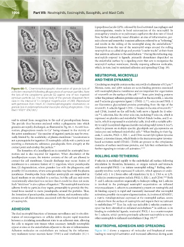
Figure 66–1. Cinemicrophotographic observation of granule lysis of Moesin, ezrin, and p205 radixin are actin-binding proteins associated
a chicken neutrophil following phagocytosis of zymosan particles. Note with neutrophil plasma membranes and are important for organization
the lysis of the cytoplasmic granule (G) against one of two ingested of microvilli on the surface of the cell. 16,17 These actin binding proteins
zymosan particles (Z). The dense body of the granule disappears from tether the primary adhesion proteins exposed on the microvilli, L-selectin
view in the interval of 5 s (original magnification ×1200). (Reproduced and P-selectin glycoprotein ligand 1 (PSGL-1). L-selectin and PSGL-1
18
with permission from Hirsch JG: Cinemicrophotographic observations on are filamentous glycosylated proteins protruding from the tips of the
granule lysis in polymorphonuclear leucocytes during phagocytosis. J Exp microvilli. E-selectin ligand 1 (ESL-1) located in the side of microvilli,
19
Med 116:827–834,1962.)
and CD44, located on the cell body, both serve a ligands for E-selec-
tin. L-selection, like the other selectins, including P-selectin, which is
20
expressed on platelets and endothelial Weibel-Palade bodies, and E-se-
said to extend from recognition to the end of pseudopodium fusion. lectin, which is expressed in endothelial cells, bind with a variable affin-
The particle thus becomes enclosed within a phagosome into which ity to sialyl fucosylated oligosaccharides including sialyl Lewis X (sLe ),
x
granules are rapidly discharged, as illustrated in Fig. 66–1. As with loco- which is present on multiple specific glycolipids and glycoproteins on
motion, phagocytosis results in Ca being released in the vicinity of leukocytes and inflamed endothelial cells. When binding to their lig-
2+
21
the active membranes. The number of ingested particles may be even- ands, L-selectin, PSGL-1, ESL-1, and CD44 recruit Syk (spleen tyrosine
6
tually limited by the availability of plasma membrane. Locomotion is kinase), a tyrosine kinase, which binds to the immunoreceptor tyrosine-
7
not a prerequisite for ingestion: If neutrophils collide with a particle not based activation motif (ITAM). ITAMs are present in the cytoplasmic
secreting a chemotactic substance, pseudopodia form abruptly at the domains of surface membrane proteins, and Syk then orchestrates the
contact point and envelop the particle. 12 further signaling to initiate cell activation. 22–25
The formation of a lamellipodium is essential for neutrophil loco-
motion and is also required for ingestion. When dissolution of the
lamellipodium occurs, the interior contents of the cell are allowed to ROLLING AND TETHERING
contact the cell membrane. Granule discharge may occur. Fusion of P-selectin is mobilized rapidly to the endothelial cell surface following
membranes is a common feature of (1) ingestion, where pseudopodia stimulation by thrombin, histamine, or oxygen radicals and interacts
fuse; (2) degranulation, where granules fuse with the phagosome; and with neutrophil PSGL-1 to initiate neutrophil rolling. Rolling subse-
21
possibly (3) locomotion, where some granules may fuse with the plasma quently involves newly expressed E-selectin, which appears on endo-
membrane. Pseudopodia form whether neutrophils are suspended in thelial cells 1 to 2 hours after cell stimulation by IL-1, TNF-α, or LPS.
liquid medium or are attached to a surface, but the cell can only move E-selectin counterreceptors include PSGL-1, ESL-1, and CD44. 19,24 Both
translationally when fixed to a surface; thus it crawls but does not swim. P- and L-selectin contribute sequentially to leukocyte rolling, but L-selectin
Such “stickiness” is also a phase of ingestion. The neutrophil membrane is involved in the prolonged neutrophil sequestration on inflamed
7
adheres firmly to particles they ingest, presumably to provide the fric- microvasculature. L-selectin is constitutively present on neutrophils and
tional force needed to move pseudopodia around the particles. Thus, its binding capacity is rapid and transiently increased after neutrophil
the formation of pseudopodia, membrane fusion, and membrane adhe- activation, possibly via receptor oligomerization. Activation of ADAM17,
siveness are all characteristics associated with the functional responses a matrix metalloproteinase expressed at the neutrophil surface, severs
of neutrophils. L-selectin from the surface of neutrophils and impairs their recruitment
to endothelium. 26,27 Thus far, only one inducible L-selectin counterrec-
28
ADHESION eptor has been identified on inflamed endothelium. In addition to its
binding to endothelial ligands, neutrophil PSGL-1 is a counterreceptor
The dual neutrophil functions of immune surveillance and in situ elim- for L-selectin, which permits previously adherent neutrophils to recruit
ination of microorganisms or cellular debris require rapid transition other neutrophils to inflamed endothelium (Chap. 19). 13,21
between a circulating nonadherent state to an adherent state, allowing
the cells to migrate into tissues when necessary. Initially neutrophils
appear at sites on the endothelium adjacent to the site of inflammation. NEUTROPHIL ADHESION AND SPREADING
Adhesion molecules on endothelium are induced by the inflamma- Figure 66–2 shows a sequence of molecular and biophysical events
tory mediators tumor necrosis factor (TNF)-α and interleukin (IL)-1. leading to neutrophil activation and increased adherence during acute
Kaushansky_chapter 66_p1005-1042.indd 1006 9/21/15 10:47 AM