Page 1056 - Williams Hematology ( PDFDrive )
P. 1056

1030  Part VII:  Neutrophils, Eosinophils, Basophils, and Mast Cells  Chapter 66:  Disorders of Neutrophil Function  1031


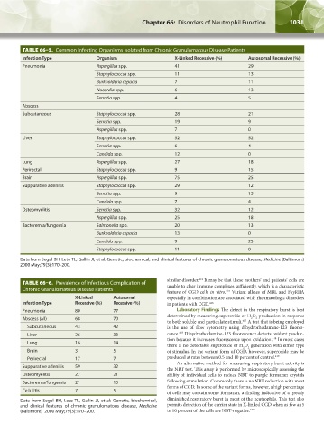
                   TABLE 66–5.  Common Infecting Organisms Isolated from Chronic Granulomatous Disease Patients
                   Infection Type             Organism                     X-Linked Recessive (%)      Autosomal Recessive (%)
                   Pneumonia                  Aspergillus spp.             41                          29
                                              Staphylococcus spp.          11                          13
                                              Burkholderia cepacia         7                           11
                                              Nocardia spp.                6                           13
                                              Serratia spp.                4                           5
                   Abscess                                                                              
                   Subcutaneous               Staphylococcus spp.          28                          21
                                              Serratia spp.                19                          9
                                              Aspergillus spp.             7                           0
                   Liver                      Staphylococcus spp.          52                          52
                                              Serratia spp.                6                           4
                                              Candida spp.                 12                          0
                   Lung                       Aspergillus spp.             27                          18
                   Perirectal                 Staphylococcus spp.          9                           15
                   Brain                      Aspergillus spp.             75                          25
                   Suppurative adenitis       Staphylococcus spp.          29                          12
                                              Serratia spp.                9                           15
                                              Candida spp.                 7                           4
                   Osteomyelitis              Serratia spp.                32                          12
                                              Aspergillus spp.             25                          18
                   Bacteremia/fungemia        Salmonella spp.              20                          13
                                              Burkholderia cepacia         13                          0
                                              Candida spp.                 9                           25
                                              Staphylococcus spp.          11                          0

                  Data from Segal BH, Leto TL, Gallin JI, et al: Genetic, biochemical, and clinical features of chronic granulomatous disease, Medicine (Baltimore)
                  2000 May;79(3):170–200.

                                                                        similar disorder.  It may be that these mothers’ and patients’ cells are
                                                                                    414
                   TABLE 66–6.  Prevalence of Infectious Complication of   unable to clear immune complexes sufficiently, which is a characteristic
                   Chronic Granulomatous Disease Patients               feature of CGD cells  in vitro.  Variant alleles of MBL and FcγRIIA
                                                                                              415
                                      X-Linked      Autosomal           especially in combination are associated with rheumatologic disorders
                   Infection Type     Recessive (%)  Recessive (%)      in patients with CGD. 416
                   Pneumonia          80            77                      Laboratory Findings The defect in the respiratory burst is best
                                                                        determined by measuring superoxide or H O  production in response
                   Abscess (all)      68            70                                                  2  2
                                                                        to both soluble and particulate stimuli.  A test that is being employed
                                                                                                    417
                     Subcutaneous     43            42                  is the use of flow cytometry using dihydrorhodamine-123 fluores-
                                                                             418
                     Liver            26            33                  cence.  Dihydrorhodamine-123 fluorescence detects oxidant produc-
                                                                        tion because it increases fluorescence upon oxidation.  In most cases
                                                                                                                418
                     Lung             16            14
                                                                        there is no detectable superoxide or H O  generation with either type
                                                                                                     2
                                                                                                      2
                     Brain            3             5                   of stimulus. In the variant form of CGD, however, superoxide may be
                     Perirectal       17            7                   produced at rates between 0.5 and 10 percent of control. 419
                                                                            An alternative method for measuring respiratory burst activity is
                   Suppurative adenitis  59         32
                                                                        the NBT test. This assay is performed by microscopically assessing the
                   Osteomyelitis      27            21                  ability of individual cells to reduce NBT to purple formazan crystals
                   Bacteremia/fungemia  21          10                  following stimulation. Commonly there is no NBT reduction with most
                                                                        forms of CGD. In some of the variant forms, however, a high percentage
                   Cellulitis         7             5                   of cells may contain some formazan, a finding indicative of a greatly
                  Data from Segal BH, Leto TL, Gallin JI, et al: Genetic, biochemical,    diminished respiratory burst in most of the neutrophils. This test also
                  and clinical features of chronic granulomatous disease,  Medicine     permits detection of the carrier state in X-linked CGD when as few as 5
                  (Baltimore)  2000 May;79(3):170–200.                  to 10 percent of the cells are NBT-negative. 420


          Kaushansky_chapter 66_p1005-1042.indd   1031                                                                  9/21/15   10:48 AM
   1051   1052   1053   1054   1055   1056   1057   1058   1059   1060   1061