
Page 1175 - Williams Hematology ( PDFDrive )
P. 1175
1150 Part IX: Lymphocytes and Plasma Cells Chapter 74: Lymphopoiesis 1151
Human
T cells
Thymus
Fetal Marrow
Fetal liver
PAS/AGM B cells
Placenta
Yolk sac
18 20 22 24 26 28 30 32 34 36 38 40 10 20 30 Birth
Circulation
Days Weeks
A
Mouse
T cells
B cells
Thymus Fetal Marrow
Fetal liver
PAS/AGM
Placenta
Yolk sac
8.0 9.0 10.0 11.0 12.0 13.0 14.0 15.0 Birth
Circulation
Days
B
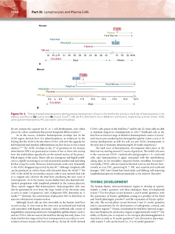
Figure 74–1. Timing of lymphohematopoiesis during prenatal development. Shown is the timeline for activity in each site of hematopoiesis in the
embryo and fetus of (A) human and (B) mouse. B and T cells are first detected in vivo in fetal liver and thymus, respectively, at times shown. AGM,
aorto-gonad-mesonephros; PAS, para-aortic splanchnopleura.
22
do not possess the capacity for B- or T-cell development, even when CD34+ cells appear in the fetal liver, and by day 32, these cells are able
placed in culture conditions that permit lymphoid differentiation. 18 to maintain long-term hematopoiesis in vitro. Erythroid cells in the
22
As in the mouse, definitive hematopoiesis develops first in the fetal liver at this later stage of definitive hematopoiesis consist of enucle-
AGM region derived from the splanchnopleura, as evidenced by the ated macrocytes producing fetal hemoglobin (globin chains α and γ). In
finding that the AGM is the site where CD34+ cells with the capacity for normal development, as with the yolk sac and AGM, hematopoiesis in
full lymphoid and myeloid differentiation are first found in the human the fetal liver is transient, disappearing by 20 weeks of gestation. 1
embryo. 18,19 The AGM develops at day 27 of gestation in the human, The final wave of hematopoietic development takes place in the
when human HSCs are generated as clusters of two or three cells arising fetal marrow, starting around 11 weeks of gestation. The initial cells seen
from the endothelium specifically on the ventral surface of the preum- in the marrow are CD15+ myeloid cells and glycophorin A+ erythroid
bilical region of the aorta. These cells are clonogenic and highly prolif- cells, and hematopoiesis is again associated with the endothelium,
23
erative, rapidly increasing to several thousand in number and spreading taking place in the medullary sinusoids before osteoblast formation.
further along the aorta. However, hematopoiesis exists only transiently Eventually, CD34+ cells are found in the fetal marrow and behave func-
17
in the AGM, disappearing entirely by day 40. Although lymphoid cells tionally as true HSC, generating B, T, NK, and myeloid and erythroid
1
can be produced in culture from cells extracted from the AGM 17,18 the lineages. HSC have found their final niche, and lifelong, self-renewing
HSC of the AGM do not produce mature cells in situ; instead their role lymphohematopoiesis resides permanently in the marrow thereafter.
is to migrate and colonize the fetal liver, producing the next wave of
hematopoiesis. As in the mouse, recent studies have also detected mul-
tipotent progenitors with lymphoid potential in the human placenta. THYMIC DEVELOPMENT
These reports suggest that hematopoietic stem/progenitor cells may The human thymic microenvironment begins to develop at approx-
also be generated de novo from the large vessels of the chorionic plate imately 4 weeks’ gestation and then undergoes three developmental
24
as early as week 5 of gestation, with multipotent HSC detectable at 15 phases. The first phase occurs between 4 and 8 weeks’ gestation, with
weeks. 20,21 However, the contribution of placental HSC to fetal liver or the appearance of thymic epithelium arising as a product of the third
25
marrow colonization remains unclear. and fourth pharyngeal pouches and the expansion of thymic epithe-
Although blood cells are first detectable in the human fetal liver lial cells. The second phase occurs between 9 and 15 weeks’ gestation
as early as day 23, they exist at this time only as erythroid and myeloid and is characterized by the development of subcapsular, cortical, and
cells associated with hepatic sinusoids. These erythroid cells consist of medullary regions. Thymic colonization by fetal liver-derived progen-
24
25
megaloblasts expressing embryonic hemoglobins (globin chains ζ and ε), itors and lymphocyte production begins at approximately week 9. The
and no CD34+ cells are seen in the fetal liver during this early phase. It is ability of thymocytes to respond to the mitogen phytohemagglutinin is
likely that this first stage of fetal liver hematopoiesis is secondary to colo- detectable as early as 10 weeks’ gestation, and alloreactive, phenotypi-
26
nization of more mature cells from the yolk sac. By day 30, AGM-derived cally mature T cells can be found by 13 to 16 weeks’ gestation. 27
Kaushansky_chapter 74_p1149-p1158.indd 1150 9/18/15 2:25 PM