
Page 1347 - Williams Hematology ( PDFDrive )
P. 1347
1322 Part X: Malignant Myeloid Diseases Chapter 86: Primary Myelofibrosis 1323
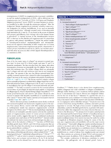
overexpression of FKBP51 in megakaryocytes in primary myelofibro- TABLE 86–1. Fibroplasia in Primary Myelofibrosis
sis and the marked predisposition of CD34+ cells to differentiate into
megakaryocytes (see “Centrality of CD34+ Cell Egress and Neoplastic I. Marrow stroma
Megakaryocytopoiesis” above). FKBP51 increases resistance to apopto- A. Increased amount of
sis, possibly by an effect through the calcineurin pathway. Thus, the 1. Total collagen (hydroxyproline) 97,101
91
designation “chronic megakaryocytic leukemia” would be a more accu- 2. Type I collagen 97–99,103
rate designation for primary myelofibrosis, if one used an internally 97–99,103
7
consistent classification. Although elevated levels of thrombopoietin 3. Type III collagen
(and interleukin [IL]-6 and IL-11) are found in the serum of patients 4. Type III procollagen 98,101,103,104
with primary myelofibrosis, their etiologic role in the human disease 5. Type IV collagen 98,105,106
92
is unresolved. A marked increase in the thrombopoietin receptor 6. Matrix metalloproteinase-14 107
MPL is observed on the platelets and megakaryocytes of a proportion 7. Bone morphogenetic protein 108
93
of patients with primary myelofibrosis. Despite the animal models 8. Laminin 98,105,109
of thrombopoietin-induced myeloproliferation and osteomyelofi- 110,111
brosis and the apparent abnormality of MPL receptor sites on human 9. Fibronectin
megakaryocytes, autonomous megakaryocyte growth, characteristic of 10. Tenascin 112
human primary myelofibrosis marrow in culture, has not been associ- 11. Vitronectin 113
ated with either an autocrine effect of MPL ligand (thrombopoietin) or 12. Microenvironment transforming growth factor-β,
114
of a mutation in MPL. basic fibroblast growth factor, and substance P 115
114
B. Decreased amount of
FIBROPLASIA 1. Collagenase 107
Four of the five major types of collagen are present in normal mar- II. Plasma
94
row: type I in bone, type III in blood vessels, and types IV and V in A. Increased concentration of
basement membranes. The fine reticulin fibers that appear after silver 1. Prolylhydroxylase 116
impregnation of marrow are principally type III collagen. They do not 2. C-terminal peptide of procollagen type I 100
stain with trichrome dyes. The thicker collagen fibers are principally 3. N-terminal peptide of procollagen type III 99,101,117,118
type I collagen and stain with trichrome dyes, but do not impregnate 4. Type IV collagen 99,109
with silver. The amount of the very fine fibrous network barely per- 5. Laminin 99,109
ceptible in normal marrow that is stained by silver impregnation tech- 110,111
niques increases in the marrow of patients with primary myelofibrosis 6. Fibronectin
95
(Table 86–1 and Fig. 86-1C). The fibrous network contains collagen 7. Hyaluronan 119
96
and occasionally progresses to include thick collagen bands that are evi-
dent with trichrome stains. Collagen types I, III, IV, and V are increased
in myelofibrosis, but type III collagen is increased uniformly and pref-
erentially. 97–104 The latter occurrence accounts for the increased plasma fibroblasts. 130–132 Platelet factor 4, also derived from megakaryocytes,
120
concentration of procollagen III aminoterminal peptide, a component inhibits collagenase and could contribute to collagen accumulation,
of collagen type III, which is cleaved during collagen biosynthesis. 96,101,102 although studies showing a poor correlation between plasma platelet
Serum prolyl-hydroxylase and marrow and plasma fibronectin also factor 4 concentration and marrow fibrosis have dampened enthusi-
increase in patients with idiopathic myelofibrosis or myelofibrosis from asm for the role of this factor. Substance P, a peptide that acts as a
133
other causes. 98,99 Several other matrix materials are increased in marrow neurotransmitter and a modulator of immune and hematopoietic func-
or plasma (Tables 86–1 and 86–2). 105–119 tions, is increased in the fibrotic marrow and colocalizes with fibronec-
Marrow fibrosis in primary myelofibrosis is most closely correlated tin. It is angiogenic and is a fibroblast mitogen. Its precise role in the
115
with increased neoplastic and dysmorphic megakaryocytes in the mar- complex interactions among fibroblasts, cytokines, and matrix protein
row. Even densely fibrotic marrow with little residual granulopoiesis deposition is not clear. The high urinary excretion of platelet-derived
or erythropoiesis usually has numerous megakaryocytes scattered calmodulin, a putative fibroblast growth factor, in patients with myelo-
throughout the fibrotic areas. 96,120 The increased pathologic emperipole- fibrosis has added this compound to the array of factors that may con-
sis (the entry of neutrophils and other marrow cells into the canalicular tribute to the fibroplasia. The plasma level of matrix metalloprotein
130
system of megakaryocytes), evident in human primary myelofibrosis III is decreased and the level of tissue inhibitor of metalloproteinase is
and in mouse models, suggests this may be an additional mechanism increased in patients with idiopathic myelofibrosis. The expression of
134
121
of α-granule injury and release of TGF-β and PDGF. Animal models matrix metalloproteinase-14 in marrow increases by nearly two orders
also indicate that marrow monocytes and macrophages may play a sub- of magnitude as fibroplasia progresses during the course of the dis-
sidiary role in the induction of fibrosis. 121–123 Secretion of PDGF, basic ease; and, megakaryocytes and endothelial cells are the major sources
107
fibroblast growth factor (bFGF), and TGF-β from monocytes that are of this protein. Neutrophil collagenase (matrix metalloproteinase-8)
part of the clone have the potential to act as myeloproliferative growth content is decreased early in the disease. Bone morphogenetic pro-
107
factors and profibrotic cytokines. 114 teins (BMPs) also are implicated as a contributory factor in fibroplasia.
The increased content of marrow collagen types I and III results BMP1, BMP6, BMP7, and BMP-receptor 2 are increased in marrow in
from release of fibroblast growth factors, which include PDGF, 123,124 epi- myelofibrosis as a result of release from megakaryocytes and stromal
126
126
dermal growth factor, endothelial cell growth factor, TGF-β, 114,127,128 cells. These proteins are activators of latent TGF-β and processors of
1
and bFGF, 114,129 each of which is present in megakaryocyte α granules. collagen precursors. In addition, TGF-β induces release of BMP6. 108
1
Other factors, such as tumor necrosis factor α, IL-1α, IL-1β, and lysyl This complex combination of alterations contributes to matrix
oxidase, which can be released from marrow cells, also can stimulate deposition. The pathogenetic role of released growth factors in
Kaushansky_chapter 86_p1319-1340.indd 1322 9/18/15 10:22 AM