
Page 1348 - Williams Hematology ( PDFDrive )
P. 1348
1322 Part X: Malignant Myeloid Diseases Chapter 86: Primary Myelofibrosis 1323
A B
C D
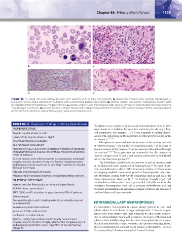
Figure 86–1. Blood film and marrow sections from patients with primary myelofibrosis. A. Blood film. Characteristic teardrop poikilocytes, a
nucleated red cell, and a segmented neutrophil with a dysmorphic nucleus are evident. B. Marrow section. Low power. Hypercellular marrow with
increased number of hypolobular megakaryocytes. C. Marrow section. Silver impregnation stain. Marked increase in argentophilic fibers representing
collagen type III (reticulin). D. Marrow section. Collagen fibrosis with extensive replacement of marrow with swirls of collagen fibers. (Reproduced with
permission from Lichtman’s Atlas of Hematology, www.accessmedicine.com.)
TABLE 86–2. Diagnostic Findings in Primary Myelofibrosis
fibroplasia is not completely understood. Generalizations from in vitro
PREFIBROTIC STAGE experiments or correlation between two variables provide only a lim-
Anemia may be absent or mild ited perspective. For example, TGF-β can stimulate or inhibit fibrob-
Leukocytosis may be absent or slight last growth, depending on the repertoire of other growth factors in the
environment.
127,128
Thrombocythemia is invariable Fibroplasia is associated with an increase in the number and size
BCR-ABL fusion gene absent of marrow sinuses, the number of endothelial cells, an increase in
135
111
Presence of JAK2, CALR, or MPL mutations indicative of diagnosis vascular volume in the marrow, and an increase in blood flow through
106
of myeloproliferative disease (one of these mutations present in the marrow. 136,137 These processes are responsible for the increase in
~90% of patients) marrow collagen types IV and V and laminin synthesized by endothelial
Cellular marrow with mild increase in granulopoiesis; increased cells in the marrow of patients. 126
megakaryocytes, clusters of very dysmorphic megakaryocytes The fibroblastic proliferation in marrow is not an intrinsic part
and megakaryocytic nuclei; no to very slight increase in reticular of the abnormal clonal expansion of hematopoiesis. In cases of pri-
138
fibers on silver stain mary myelofibrosis in which G6PD isoenzyme studies or chromosome
Palpable splenomegaly infrequent karyotyping establish monoclonal growth of hematopoietic cells, mar-
Absent or slight anisopoikilocytosis including teardrop red cells row fibroblasts contain both G6PD isoenzymes and do not share the
139
FULLY DEVELOPED STAGE clonal chromosome abnormality. The findings strongly imply that
the fibroblasts differentiate from a primordial cell different from the
Marrow reticulin fibrosis plus or minus collagen fibrosis neoplastic hematopoietic stem cell in primary myelofibrosis and that
BCR-ABL fusion gene absent fibroblast proliferation and enhanced collagen synthesis are secondary
JAK2, CALR, or MPL mutation in approximately 90% of patients results of abnormal hematopoiesis.
Splenomegaly
Anisopoikilocytosis with teardrop red cells in virtually every oil
immersion field EXTRAMEDULLARY HEMATOPOIESIS
Immature myeloid cells in blood Extramedullary hemopoiesis is almost always present in liver and
Increased CD34+ cells in blood spleen, where it contributes to organ enlargement. 8–10 Escape of pro-
Nucleated red cells in blood genitor cells from marrow and their lodgment in other organs contrib-
utes to extramedullary blood cell formation. Reversion of the liver and
Marrow usually hypercellular but invariably has increased spleen to their fetal hematopoietic functions (metaplasia) is not a major
megakaryocytes, clusters of highly dysmorphic megakaryocytes, factor in extramedullary hematopoiesis, and quantitatively significant,
and megakaryocyte bare nuclei regardless of overall marrow
cellularity effective hematopoiesis does not occur outside of the marrow (see also
“Extramedullary (Fibrohematopoietic) Tumors” below).
Kaushansky_chapter 86_p1319-1340.indd 1323 9/18/15 10:23 AM