
Page 1350 - Williams Hematology ( PDFDrive )
P. 1350
1324 Part X: Malignant Myeloid Diseases Chapter 86: Primary Myelofibrosis 1325
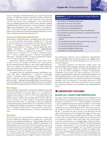
factors. Pulmonary arterial hypertension, also, may be the principal TABLE 86–3. Serum, Urine, and Bone Changes Reflecting
224
problem. 225,226 Although as many as one-third of patients with primary 243,244
myelofibrosis have an elevated systolic pulmonary artery pressures Osteosclerosis
(>35 torr), the fraction that is symptomatic is very small. Elevated • Increased serum alkaline phosphatase
vascular endothelial growth factor (VEGF) levels, elevated circulating • Increased serum bone GLA-protein
endothelial cells, and elevated marrow microvessel density in patients • Increased serum carboxytelopeptidase
suggest that proangiogenic factors may contribute to the hyperten- • Increased urinary deoxypyridinoline
sion. Contrariwise, secondary myelofibrosis with polyclonal hemato-
227
poiesis and normal blood CD34 cell concentrations frequently occurs in • Increased bone density by dual-energy x-ray absorption
patients with primary pulmonary hypertension. 228 • Increased bone density by quantitative computed tomography
• Histomorphometry
Immune and Inflammatory Manifestations • Increased percentage of cancellous bone volume to tissue
Abnormalities of humoral immune mechanisms have been observed volume
in up to half of patients with primary myelofibrosis. 229–234 The array • Increased bone formation and resorption (high turnover)
of immune products and events reported includes anti–red cell anti- • Increased trabecular plate thickness
bodies, 233–237 antiplatelet antibodies, 238,239 antinuclear antibodies, 229,230,234
240
elevated plasma-soluble IL-2 receptor, anti-Gal (galactoside deter- • Increased percentage of woven bone volume
241
minants) antibodies, anti–γ-globulins, 229,230,234 antiphospholipid • Increased percentage of fibrous area
antibodies, 234,242 antitissue or organ-specific antibodies, 231,233 and cir- • No evidence of mineralization defect
culating immune complexes, 234,243–245 as well as complement activa-
tion, 234,246 immune complex deposition, interstitial immunoglobulin
231
231
deposition, increased numbers of marrow plasmacytoid lympho- The cardiovascular risk factor, such as hypertension, hypercholester-
cytes, 231,243 and development of amyloidosis. 244–247 olemia, or smoking, further increase thrombotic risk. Multiple throm-
Inflammatory cytokines, including IL-1β, IL-6, IL-8, tumor necro- botic episodes may occur; and, the thrombotic event may occur at or
sis factor (TNF)-α, TNF receptor II (TNFRII), and C-reactive protein just before diagnosis.
also are markedly elevated and play a role in the constitutional symp- Noncirrhotic splanchnic vein thrombosis includes hepatic vein
248
toms seen in patients with progressive disease, which explains the thrombosis (Budd-Chiari syndrome) and portal vein thrombosis, which
often rapid, symptomatic improvement in patients treated with JAK2 may occur with minimal evidence of a clonal myeloproliferative dis-
inhibitors before splenic size is reduced. ease. In the past marrow examination or evidence of erythropoietin-
Occasional reports of nonclonal secondary myelofibrosis asso- independent colony growth was used to determine if an occult or
255
ciated with lupus erythematosus, 249–254 vasculitis, polyarteritis incipient myeloproliferative disease may underlie the thrombosis. Now
257
256
nodosa, 234,255 ulcerative colitis, scleroderma, biliary cirrhosis, 237,258,259 JAK2 mutational analysis can be done and is positive in 35 percent of
Sjögren syndrome, and acute reversible myelofibrosis responsive to seemingly idiopathic hepatic vein thrombosis and 25 percent seemingly
260
glucocorticoids, although fundamentally different processes from pri- idiopathic portal vein thrombosis. Presumably, future use of CALR
261
276
mary myelofibrosis, have raised the possibility that immune mechanisms gene mutational analysis will increase the proportion of cases of hepatic
play a role in the development of marrow fibrosis in some circumstances. vein thrombosis indicative of an underlying occult myeloproliferative
disease.
Bone Changes
A large proportion of patients have osteosclerosis at diagnosis or develop
osteosclerosis during the course of the disease, 11–15,262–265 as reflected by LABORATORY FEATURES
increased bone density on imaging studies and histomorphometric
analysis of a bone biopsy (Table 86–3). 263–268 The proximal femur and BLOOD CELL COUNTS AND MORPHOLOGY
humerus, pelvis, vertebrae, ribs, and skull may be involved. MRI can The range of values for blood cell counts at the time of diagnosis is very
uncover evidence of new bone formation and periosteal thickening. broad. Normocytic–normochromic anemia is present in most, but not
Lumbar spine dual-energy x-ray absorption studies and quantitative all, patients (see Table 86–2). 8–10,273–280 Mean hemoglobin concentration
computed tomography provide evidence for increased bone formation, in a series of patients at diagnosis was approximately 9 to 12 g/dL (range:
bone thickening, and higher proportions of cancellous and of woven 4–20 g/dL). 8–16,279,280 Anisocytosis and poikilocytosis are a constant
270
bone. 268,269 Osteolytic lesions are rare and may reflect a myeloid sar- finding. In all cases, teardrop-shaped red cells (dacryocytes) are pres-
coma. Periostitis, although infrequent, can lead to debilitating bone ent in sufficient number to be found in every oil immersion field (see
271
pain. 272 Fig. 86–1). Nucleated red cells are present in the blood film of most
patients and average 2 percent of nucleated cells (range: 0 to 30 percent).
Thrombosis The percentage of reticulocytes is mildly increased but may vary widely
The risk of arterial and venous thrombosis is elevated in patients with in a given case. A decreased blood hemoglobin may be attributed in part
primary myelofibrosis, although not to the degree seen in polycythe- to the expansion of plasma volume and a higher than normal propor-
273
mia vera or essential thrombocythemia. Approximately 10 percent of tion of the red cell volume in an enlarged spleen. Ineffective erythro-
277
patients with myelofibrosis will develop a significant thrombotic event poiesis can result in a decrease in red cell mass. Erythroid hypoplasia
during the first 4 years of the disease. The two principal risk factors are is present in many patients. 281,282 In some patients, hemolysis may be
274
an elevated leukocyte count and age, but not platelet count. In a large prominent, and polychromatophilia and very elevated reticulocyte
multicenter study of 707 patients with primary myelofibrosis, thrombo- counts can occur. 278,279 The antiglobulin (Coombs) test usually is nega-
ses occurred in 7.2 percent of patients over the period of observation, tive, but red cell autoantibodies can develop and lead to immune-medi-
or 1.8 percent patient-years. The combination of the JAK2 mutation, ated hemolysis, 234–236,283 which rarely has been a presenting finding of the
275
236
leukocytosis, and age predicted the highest incidence of thrombosis. disease. Occasional patients have had a positive acid hemolysis and
Kaushansky_chapter 86_p1319-1340.indd 1325 9/18/15 10:23 AM