
Page 1375 - Williams Hematology ( PDFDrive )
P. 1375
1350 Part X: Malignant Myeloid Diseases Chapter 87: Myelodysplastic Syndromes 1351
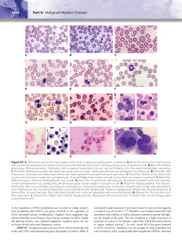
A B C
D E F
G H I
J K L
Figure 87–3. Blood and marrow films from patients with clonal cytopenias (myelodysplastic syndromes). A. Blood film. Anisocytosis. Poikilocytosis
with occasional fragmented cells. Marked anisochromia with marked hypochromia, mild hypochromia and normochromic cells. B. Blood film. Marked
anisocytosis. Mild anisochromia. Poikilocytes with occasional fragmented cells and oval and elliptical cells. Two polychromatophilic macrocytes.
C. Blood film. Striking anisocytosis with giant macrocytes and microcytes. Poikilocytes with tiny red cell fragment and elliptocyte. D. Blood film. Mild
anisocytosis. Ovalocytes and elliptocytes. Dacryocyte. Hyposegmented neutrophil with poor granulation. E. Blood film. Marked anisocytosis (mac-
rocytes and microcytes). Ovalocytes and elliptocytes. Acquired Pelger-Hüet nuclear anomaly (classic pince-nez shape) in neutrophil. F. Blood film.
Mild anisocytosis. Abnormal neutrophil with ring nucleus. G. Blood film. Anisochromia. Stomatocytes. Abnormal neutrophil nuclei with hyperlob-
ulation and hyperchromatic staining. Note abnormal elongated nuclear bridge in neutrophil on left. H. Blood film. Atypical platelets. Two macroth-
rombocytes with excess cytoplasm and atypical central granules. Anisocytosis (conspicuous microcytes). Anisochromia (conspicuous hypochromic
cells). Poikilocytosis with occasional fragmented red cells. I. Marrow film. Wright stain. Trilobed megakaryocyte. Wright stain. Macroerythroblasts. J.
Marrow films. Prussian blue stain. Ring sideroblasts. Wright stain. Erythroid hyperplasia with macroerythroblasts. K. Marrow film. Prussian blue stain.
Ring sideroblasts. L. Marrow film. Wright stain. Trilobed megakaryocytes. (Reproduced with permission from Lichtman’s Atlas of Hematology, www.
accessmedicine.com.)
in the regulation of DNA methylation are mutated in a large propor- is mutated in approximately 15 percent of cases in a pattern that suggests
tion of patients with MDS as are genes involved in the regulation of a resultant loss of function. 149,150 Mutations can include frameshifts and
DNA-associated histone modifications. Together these epigenetic reg- premature stop codons, as well as missense mutations spread through-
ulators form the second largest class of genes mutated in MDS. Unlike out the length of the gene. The one exception is a high frequency of
the splicing factors, most mutated epigenetic regulator genes are not missense mutations at the hotspot codon 882, which have been shown
exclusive of each other and frequently coexist. to impair catalytic activity. As with nearly all of the genes mutated
151
DNMT3A This gene encodes a de novo DNA methyltransferase and in MDS, DNMT3A mutations are not unique to these disorders and
is the only DNA methyltransferase gene frequently mutated in MDS. It can be found in AML, myeloproliferative neoplasms (MPNs), and even
Kaushansky_chapter 87_p1341-1372.indd 1350 9/21/15 11:05 AM