
Page 1399 - Williams Hematology ( PDFDrive )
P. 1399
1374 Part X: Malignant Myeloid Diseases Chapter 88: Acute Myelogenous Leukemia 1375
identification of individual chromosomes and the point at which they TABLE 88–1. Conditions Predisposing to Development of
break in the formation of a translocation, inversion, or deletion. This
technologic advance unleashed the power of cancer cytogenetics and Acute Myelogenous Leukemia
initiated an era of leukemia study based not solely on the appearance Environmental factors
of cells under the microscope (phenotype) but also by their chromoso- Radiation 14,15
8
mal or genetic abnormality (genotype). The completion of the human Benzene 16–18
genome project further enhanced the specificity of the identification of Alkylating agents, topoisomerase II inhibitors, and other
9
gene alterations. These advances permitted (1) more precise under- cytotoxic drugs 20–22
standing of the molecular pathology of specific leukemia subtypes, Tobacco smoke 19,24,25
(2) improvement of diagnostic and prognostic methods for the study Acquired diseases
of AML, and (3) identification of molecular targets for therapy.
The introduction to the clinic by Holland, Ellison, and colleagues Clonal myeloid diseases
10
of arabinosyl cytosine (cytarabine) in the late 1960s as the first potent Chronic myelogenous leukemias (CML, CMML, CNL, etc.) (Chap. 89)
drug for treatment of AML, followed by their introduction of the com- Primary myelofibrosis (Chap. 86)
bination of 7 days of cytosine arabinoside and 3 days of daunorubicin Essential thrombocythemia (Chap. 85)
in the early 1970s (the “7 plus 3 regimen”) opened the era of effective Polycythemia vera (Chap. 84)
11
therapy for AML. This drug combination or its congeners remains the Clonal cytopenias (Chap. 87)
mainstay of treatment over 4 decades later. The description of alloge- Oligoblastic myelogenous leukemia(Chap.87)
12
neic marrow (stem cell) transplantation as a curative therapy for AML Paroxysmal nocturnal hemoglobinuria (Chap. 40)
13
by Thomas and colleagues in 1977 ushered in the era of hematopoietic Other hematopoietic disorders
stem cell (HSC) transplantation as a modality to cure eligible patients
with AML. Aplastic anemia (Chap. 35)
Eosinophilic fasciitis (Chap. 87)
Myeloma (Chap. 107) 31,32
ETIOLOGY AND PATHOGENESIS Other disorders
Human immunodeficiency virus infection 32
ENVIRONMENTAL FACTORS Langerhans cell histiocytosis 33,34
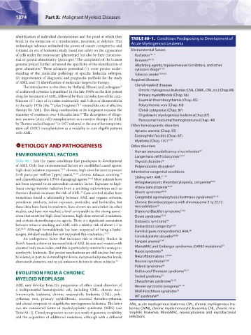
Table 88–1 lists the major conditions that predispose to development Thyroid disorders 35
of AML. Only four environmental factors are established causal agents: Polyendocrine disorders 36
high-dose radiation exposure, 14,15 chronic, high-dose benzene exposure Inherited or congenital conditions
(≥40 parts per million [ppm]-years), 16–18 chronic tobacco smoking, 37–39
19
and chemotherapeutic (DNA-damaging) agents. 20–22 Most patients have Sibling with AML
not been exposed to an antecedent causative factor. Exposure to high- Amegakaryocytic thrombocytopenia, congenital 40,41
linear energy transfer radiation from α-emitting radioisotopes such as Ataxia-pancytopenia 42,43
thorium dioxide increases the risk of AML. Case-control studies have Bloom syndrome 44,45
23
sometimes found a relationship between AML and organic solvents, Congenital agranulocytosis (Kostmann syndrome) 46–49
petroleum products, radon exposure, pesticides, and herbicides, but Chronic thrombocytopenia with chromosome 21q 22.12
these data have been inconsistent, have shown no association in other microdeletion 50
studies, and have not reached a level comparable to the strong associ- Diamond-Blackfan syndrome 51,52
ation that exists for high-dose benzene, high-dose external irradiation, Down syndrome 53,54
and certain chemotherapeutic agents. There is a significant association Dubowitz syndrome 55
between tobacco smoking and AML with a relative risk of about 1.5 to Dyskeratosis congenita 56,57
2.0. 24,25 Although formaldehyde has been suspected of being a leuke- Familial (pure, nonsyndromic) AML 58
mogen, detailed analysis has not supported this contention. 26,27 59,60
An endogenous factor that increases risk is obesity. Studies in Familial platelet disorder
61,62
North America show an increased risk of AML in men and women with Fanconi anemia
elevated body mass index, and this is particularly notable for acute pro- MonoMAC and Emberger syndromes (GATA2 mutations) 63
myelocytic leukemia. The precise mechanisms are still unclear but may Naxos syndrome 64
be related, in part, to elevated leptin levels, decreased adiponectin levels, Neurofibromatosis 1 65,66
shortened telomeres, and as yet unknown factors in obese subjects. 28 Noonan syndrome 67,68
Poland syndrome 69
EVOLUTION FROM A CHRONIC Rothmund-Thomson syndrome 70,71
72
MYELOID NEOPLASM Seckel syndrome 73–75
Shwachman syndrome
AML may develop from the progression of other clonal disorders of Werner syndrome (progeria) 76–78
a multipotential hematopoietic cell, including CML, chronic mye- Wolf-Hirschhorn syndrome 79
lomonocytic leukemia, chronic neutrophilic leukemia (CNL), poly- WT syndrome 80
cythemia vera, primary myelofibrosis, essential thrombocythemia,
and clonal cytopenia or oligoblastic myelogenous leukemia. The latter AML, acute myelogenous leukemia; CML, chronic myelogenous leu-
two are considered forms of myelodysplastic syndrome (MDS) (see kemia; CMML, chronic myelomonocytic leukemia; CNL, chronic neu-
Table 88–1). Clonal progression occurs as a result of genomic instability trophilic leukemia; MonoMAC, monocytopenia and mycobacterial
and the acquisition of additional mutations, although with a different infections.
Kaushansky_chapter 88_p1373-1436.indd 1374 9/21/15 11:00 AM