
Page 1652 - Williams Hematology ( PDFDrive )
P. 1652
1626 Part XI: Malignant Lymphoid Diseases Chapter 98: Diffuse Large B-Cell Lymphoma and Related Diseases 1627
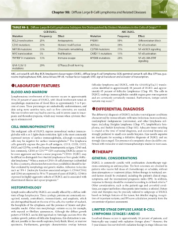
TABLE 98–2. Diffuse Large B-Cell Lymphoma Subtypes Are Distinguished By Distinct Mutations in the Cells of Origin 19–23
GCB DLBCL ABC DLBCL
Mutation Frequency Effect Mutation Frequency Effect
BCL2 translocation 25% Antiapoptotic PRDM1 50% Differentiation block
EZH2 mutations 22% Histone modification A20 loss 20% NF-κB activation
MEF2B mutations 22% Chromatin remodeling CD79B mutations 21% NF-κB/BCR signaling
MYC translocation 5% Proliferation CARD 11 mutations 11% NF-κB activation
TNFRSF14 mutations 13% Immune escape MYD88 mutations 29% NF-κB /JAK-STAT
signaling
GNA 12 & 13 29% GTPases; B-cell homing
mutations
ABC, activated B-cell–like; BCR, breakpoint cluster region; DLBCL, diffuse large B-cell lymphoma; GCB, germinal center B-cell–like; GTPase, gua-
nosine triphosphatase; KAK, Janus kinase; NF-κB, nuclear factor kappaB; STAT, signal transducer and activator of transcription.
LABORATORY FEATURES follicular lymphoma and DLBCL, with the t(14;18)(q32;q21) translo-
cation identified in approximately 20 percent of DLBCL and approx-
BLOOD AND MARROW imately 85 percent of follicular lymphomas (Chap. 99). The cells in
Lymphomatous involvement of the marrow occurs in approximately DLBCL undergo immunoglobulin variable-region gene rearrangement
and are commonly somatically mutated. Furthermore, isotype switch
10 to 20 percent of cases of DLBCL, with blood involvement noted on variants may occur. 32
morphologic examination of blood films in approximately 3 to 8 per-
cent of cases. These percentages are undoubtedly underestimates, and
data using more sensitive tests, such as flow cytometry, are needed. DIFFERENTIAL DIAGNOSIS
Marrow involvement may lead to anemia, and in severe cases to leuco-
penia and thrombocytopenia, which may worsen when cytotoxic ther- The differential diagnosis of DLBCL includes nonmalignant conditions
apy is administered. characterized by immunoblastic infiltrates (infectious mononucleosis),
nonlymphoid malignancies (carcinoma), and other lymphoma sub-
types, including Hodgkin lymphoma (Chap. 97), lymphoblastic lym-
CELL IMMUNOPHENOTYPE phoma, and Burkitt lymphoma (Chap 102). Adequate tissue sampling
The malignant cells of DLBCL express monoclonal surface immuno- is crucial at the time of initial diagnosis, and excisional biopsies are
globulin with κ or λ light-chain restriction. IgM is the most commonly strongly preferred to small core needle biopsies. Fine-needle aspirates
expressed isotype of surface immunoglobulin, although occasionally are inadequate for securing a definitive diagnosis of DLBCL and are
cells may be negative for surface immunoglobulin. The lymphoma strongly discouraged. The presence of a neoplastic clone should be con-
27
cells generally express the pan–B-cell antigens, CD19, CD20, CD22, firmed with molecular or and immunophenotypic studies in most cases.
PAX5, and CD79a, as well as the pan-hematopoietic antigen, CD45 and,
less commonly, CD10 or CD5. 27,28 CD5-expressing DLBCLs appear to THERAPY
be more aggressive and have a worse prognosis. CD10+ DLBCL may
29
be difficult to distinguish from Burkitt lymphoma or from grade 3 follic- GENERAL CONSIDERATIONS
ular lymphoma. When a mature CD10+ B-cell phenotype is identified
30
by flow cytometry, distinction between these possibilities should be fur- DLBCL is commonly curable with combination chemotherapy regi-
ther evaluated by morphology and genetic studies. Adhesion molecules mens containing an anthracycline. The best outcomes are obtained in
such as LFA-1 (leukocyte function-associated antigen-1; CD16/CD18) patients who receive full doses of chemotherapy on schedule, without
and CD44 are expressed in 50 to 75 percent of cases of DLBCL. CD44 is dose attenuations or treatment delays. Before therapy is instituted, sev-
expressed in highly aggressive subsets of DLBCL and is associated with eral factors should be evaluated, including the patient’s clinical stage,
disseminated disease and a poor prognosis. 31 symptoms, and the international prognostic index (IPI). In addition,
response to therapy should be evaluated according to defined criteria.
33
Other considerations, such as the patient’s age and comorbid condi-
HISTOPATHOLOGY tions, are important before a therapeutic intervention is selected. Future
Lymph nodes affected by DLBCL are usually effaced by a diffuse infil- trials and therapies may be precisely tailored to subgroups of DLBCL
trate of large lymphocytes. Three cytologic patterns are commonly rec- based on biologic features of the tumor, but detailed staging, recogni-
ognized, namely, centroblastic, immunoblastic, and anaplastic, which tion of important variants, and IPI score calculation presently form the
are distinguished based on the size of the cells, the number of nucleoli, cornerstone of patient assessment.
the basophilia of the cytoplasm, and the presence of bizarre and pleo-
morphic nuclei. Other rare morphologic variants occur, for example, LIMITED STAGE DIFFUSE LARGE B-CELL
with myxoid or fibrillary appearances. Although the diffuse growth
pattern of DLBCL can be distinguished on histologic sections from the LYMPHOMA (STAGES I AND II)
nodular growth pattern of follicular lymphoma, this distinction is usu- Localized disease occurs in approximately 30 percent of patients, and
ally not possible in fine-needle aspirates, body fluids, blood, or marrow historically was treated with radiation therapy alone. However, the
34
specimens. Furthermore, genotypic characteristics overlap between 5-year disease-free survival with radiation therapy in stage I disease was
Kaushansky_chapter 98_p1625-1640.indd 1627 9/18/15 11:42 PM